Japanese
English
臨床実験
硝子体の細隙灯顕微鏡的所見—第四篇 硝子体ヘルニア
The slit-lamp microscopic findings of the Vitreous body.
陳昆 暁
1
K. Chin
1
1台湾呉基福眼科研究所
1Go Kifuku Research Institute of Opthalmology
pp.451-462
発行日 1956年3月15日
Published Date 1956/3/15
DOI https://doi.org/10.11477/mf.1410205655
- 有料閲覧
- Abstract 文献概要
- 1ページ目 Look Inside
第1章 緒論
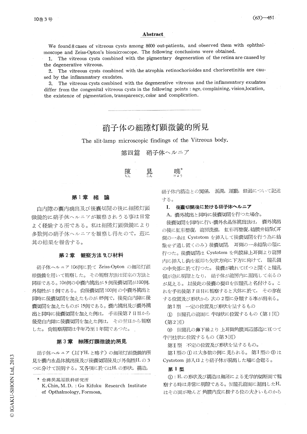
白内障の嚢内摘出及び後嚢切開の後に細隙灯顕微鏡的に硝子体ヘルニアが観察されうる事は日常よく経験する所である。私は細隙灯顕微鏡により多数例の硝子体ヘルニアを観察し得たので,茲に其の結果を報告する。
The observation of the vitreous hernias caused by intracapsular (5 cases) and extracapsular (100 cases) and trauma (1 case) with Zeiss-Opton's biomicroscope indicates the following con-clusions.
1. The vitreous hernias can be devided into two forms, one is complex hernia combined with ruptue of the anterior hyaloid membrane, the other is simple hernia without rupture of the anterior hyaloid membranc.
2. The complex hernias combined with rupture of anterior hyaloid membrane are devided into typical hernia and untypical hernia according to the existency of vitreous liquifaction.

Copyright © 1956, Igaku-Shoin Ltd. All rights reserved.


