Japanese
English
- 有料閲覧
- Abstract 文献概要
- 1ページ目 Look Inside
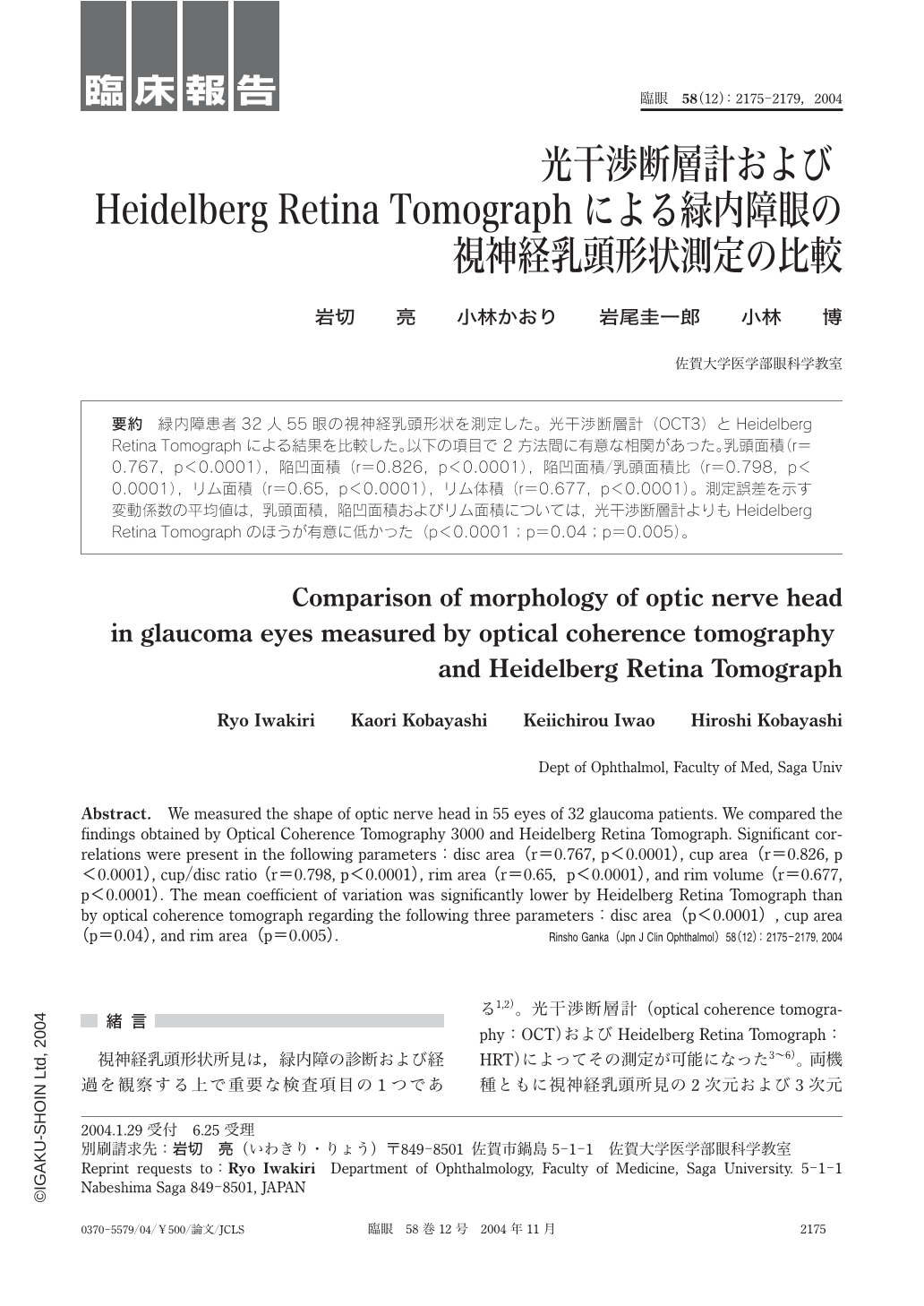
緑内障患者32人55眼の視神経乳頭形状を測定した。光干渉断層計(OCT3)とHeidelberg Retina Tomographによる結果を比較した。以下の項目で2方法間に有意な相関があった。乳頭面積(r=0.767,p<0.0001),陥凹面積(r=0.826,p<0.0001),陥凹面積/乳頭面積比(r=0.798,p<0.0001),リム面積(r=0.65,p<0.0001),リム体積(r=0.677,p<0.0001)。測定誤差を示す変動係数の平均値は,乳頭面積,陥凹面積およびリム面積については,光干渉断層計よりもHeidelberg Retina Tomographのほうが有意に低かった(p<0.0001;p=0.04;p=0.005)。
We measured the shape of optic nerve head in 55 eyes of 32glaucoma patients. We compared the findings obtained by Optical Coherence Tomography 3000 and Heidelberg Retina Tomograph. Significant correlations were present in the following parameters:disc area(r=0.767,p<0.0001),cup area(r=0.826,p<0.0001),cup/disc ratio(r=0.798,p<0.0001),rim area(r=0.65,p<0.0001),and rim volume(r=0.677,p<0.0001). The mean coefficient of variation was significantly lower by Heidelberg Retina Tomograph than by optical coherence tomograph regarding the following three parameters:disc area(p<0.0001),cup area(p=0.04),and rim area(p=0.005).

Copyright © 2004, Igaku-Shoin Ltd. All rights reserved.


