Japanese
English
- 有料閲覧
- Abstract 文献概要
- 1ページ目 Look Inside
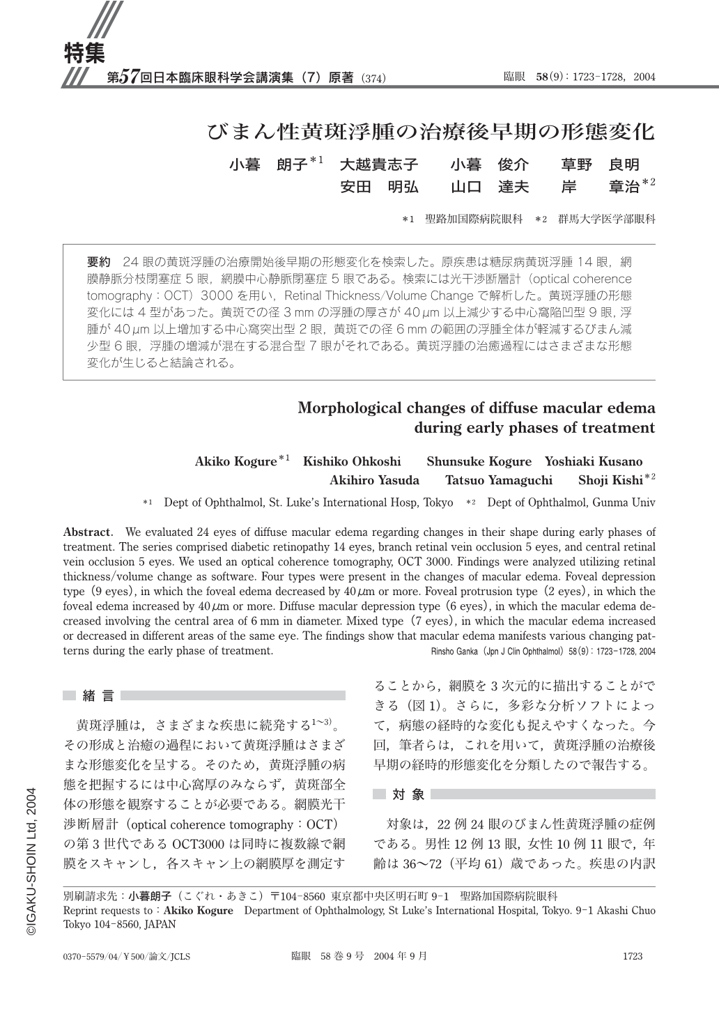
24眼の黄斑浮腫の治療開始後早期の形態変化を検索した。原疾患は糖尿病黄斑浮腫14眼,網膜静脈分枝閉塞症5眼,網膜中心静脈閉塞症5眼である。検索には光干渉断層計(optical coherence tomography:OCT)3000を用い,Retinal Thickness/Volume Changeで解析した。黄斑浮腫の形態変化には4型があった。黄斑での径3mmの浮腫の厚さが40μm以上減少する中心窩陥凹型9眼,浮腫が40μm以上増加する中心窩突出型2眼,黄斑での径6mmの範囲の浮腫全体が軽減するびまん減少型6眼,浮腫の増減が混在する混合型7眼がそれである。黄斑浮腫の治癒過程にはさまざまな形態変化が生じると結論される。
We evaluated 24 eyes of diffuse macular edema regarding changes in their shape during early phases of treatment. The series comprised diabetic retinopathy 14 eyes,branch retinal vein occlusion 5 eyes,and central retinal vein occlusion 5 eyes. We used an optical coherence tomography,OCT 3000. Findings were analyzed utilizing retinal thickness/volume change as software. Four types were present in the changes of macular edema. Foveal depression type(9 eyes),in which the foveal edema decreased by 40μm or more. Foveal protrusion type(2 eyes),in which the foveal edema increased by 40μm or more. Diffuse macular depression type(6 eyes),in which the macular edema decreased involving the central area of 6mm in diameter. Mixed type(7 eyes),in which the macular edema increased or decreased in different areas of the same eye. The findings show that macular edema manifests various changing patterns during the early phase of treatment.

Copyright © 2004, Igaku-Shoin Ltd. All rights reserved.


