Japanese
English
- 有料閲覧
- Abstract 文献概要
- 1ページ目 Look Inside
- 参考文献 Reference
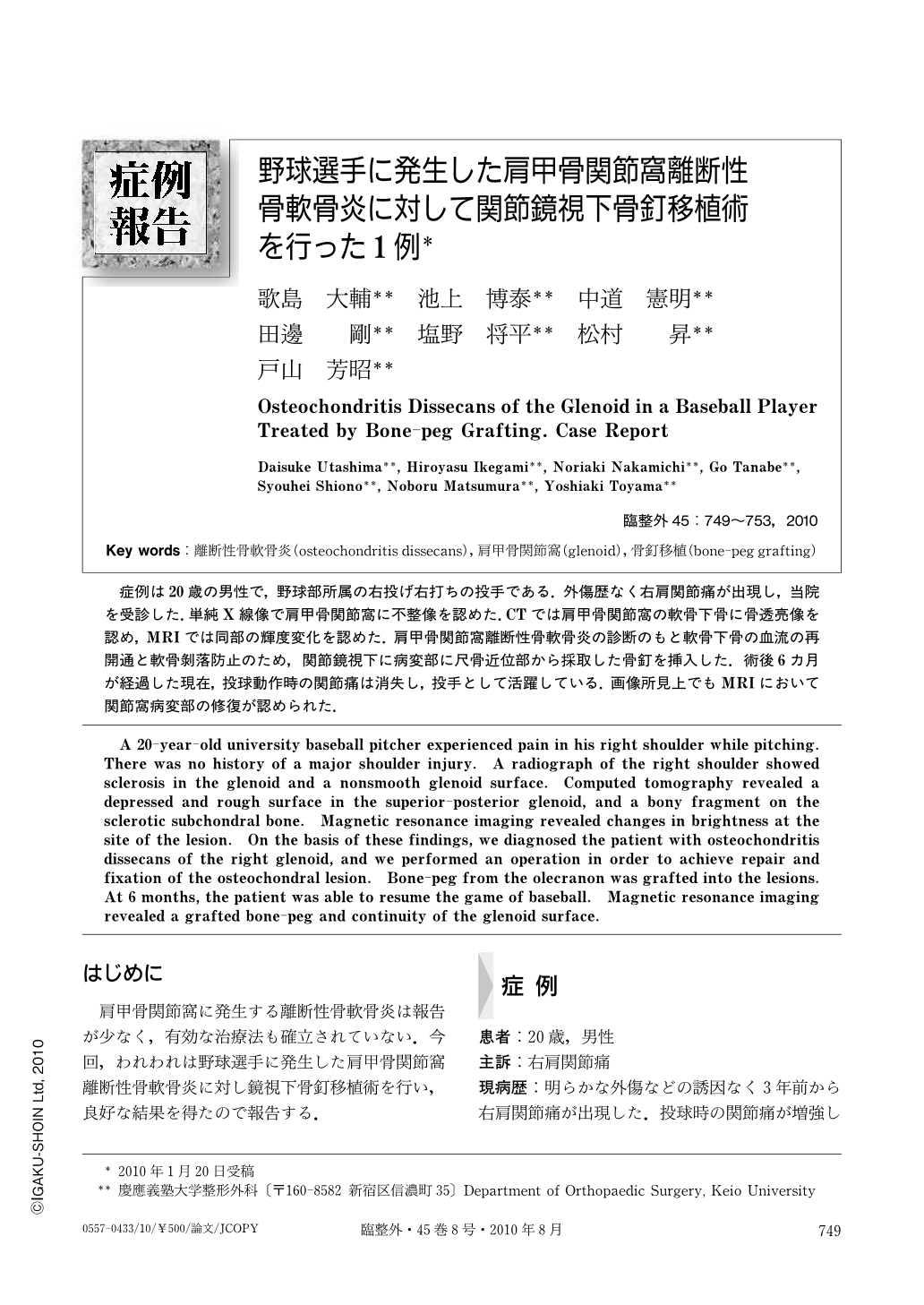
症例は20歳の男性で,野球部所属の右投げ右打ちの投手である.外傷歴なく右肩関節痛が出現し,当院を受診した.単純X線像で肩甲骨関節窩に不整像を認めた.CTでは肩甲骨関節窩の軟骨下骨に骨透亮像を認め,MRIでは同部の輝度変化を認めた.肩甲骨関節窩離断性骨軟骨炎の診断のもと軟骨下骨の血流の再開通と軟骨はく落防止のため,関節鏡視下に病変部に尺骨近位部から採取した骨釘を挿入した.術後6カ月が経過した現在,投球動作時の関節痛は消失し,投手として活躍している.画像所見上でもMRIにおいて関節窩病変部の修復が認められた.
A 20-year-old university baseball pitcher experienced pain in his right shoulder while pitching. There was no history of a major shoulder injury. A radiograph of the right shoulder showed sclerosis in the glenoid and a nonsmooth glenoid surface. Computed tomography revealed a depressed and rough surface in the superior-posterior glenoid, and a bony fragment on the sclerotic subchondral bone. Magnetic resonance imaging revealed changes in brightness at the site of the lesion. On the basis of these findings, we diagnosed the patient with osteochondritis dissecans of the right glenoid, and we performed an operation in order to achieve repair and fixation of the osteochondral lesion. Bone-peg from the olecranon was grafted into the lesions. At 6 months, the patient was able to resume the game of baseball. Magnetic resonance imaging revealed a grafted bone-peg and continuity of the glenoid surface.

Copyright © 2010, Igaku-Shoin Ltd. All rights reserved.


