Japanese
English
- 有料閲覧
- Abstract 文献概要
- 1ページ目 Look Inside
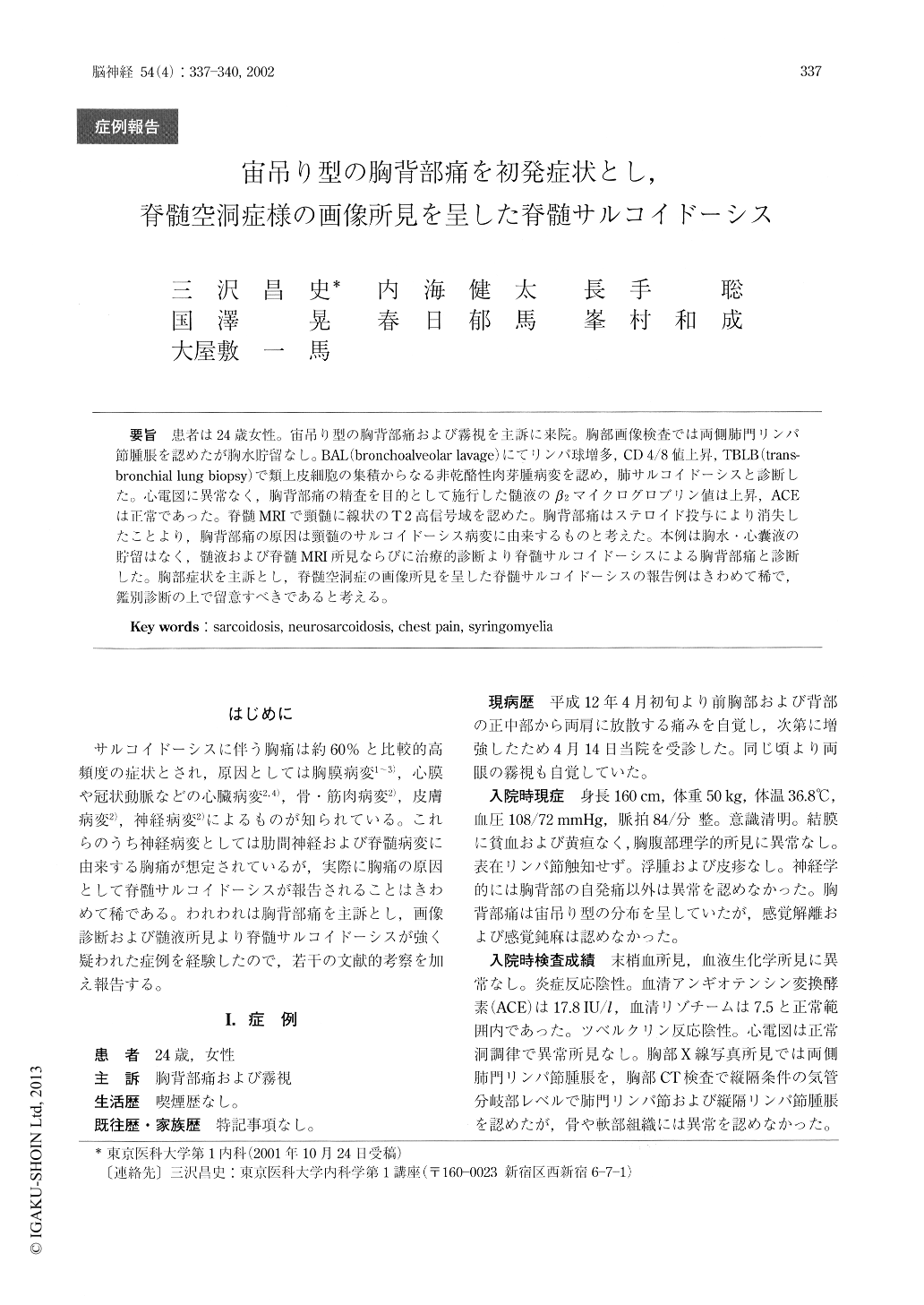
患者は24歳女性。宙吊り型の胸背部痛および霧視を主訴に来院。胸部画像検査では両側肺門リンパ節腫脹を認めたが胸水貯留なし。BAL(bronchoalveolar lavage)にてリンパ球増多,CD4/8値上昇,TBLB(trans-bronchial lung biopsy)で類上皮細胞の集積からなる非乾酪性肉芽腫病変を認め,肺サルコイドーシスと診断した。心電図に異常なく,胸背部痛の精査を目的として施行した髄液のβ2マイクログロブリン値は上昇,ACEは正常であった。脊髄MRIで頸髄に線状のT2高信号域を認めた。胸背部痛はステロイド投与により消失したことより,胸背部痛の原因は頸髄のサルコイドーシス病変に由来するものと考えた。本例は胸水・心嚢液の貯留はなく,髄液および脊髄MRI所見ならびに治療的診断より脊髄サルコイドーシスによる胸背部痛と診断した。胸部症状を主訴とし,脊髄空洞症の画像所見を呈した脊髄サルコイドーシスの報告例はきわめて稀で,鑑別診断の上で留意すべきであると考える。
The patient was a 24-year-old female complaining of bell-shaped chest and back pain with visual distur-bance. Chest X-ray showed bilateral hilar lymphade-nopathy without the presence of pleural effusion. Bronchoalveolar fluid showed lymphocytosis with an elevated CD4/CD8 ratio. Transbronchial lung biopsy demonstrated a non-caserous granulomatous lesion with an accumulation of epitheloid cells, suggesting lung sarcoidosis. No abnormality of electrocardiogram was detectable, and spinal tap for examination of chest and back pain demonstrated on elevated level of β2- microglobulin, and a normal angiotensin converting enzyme level.

Copyright © 2002, Igaku-Shoin Ltd. All rights reserved.


