Japanese
English
- 有料閲覧
- Abstract 文献概要
- 1ページ目 Look Inside
- 参考文献 Reference
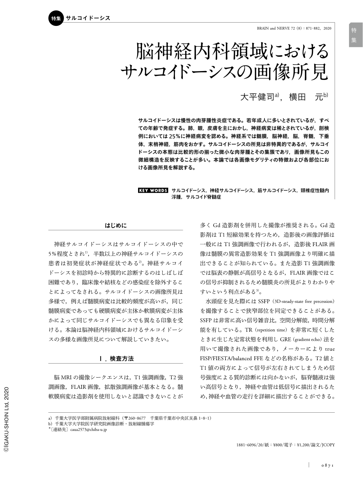
サルコイドーシスは慢性の肉芽腫性炎症である。若年成人に多いとされているが,すべての年齢で発症する。肺,眼,皮膚を主におかし,神経病変は稀とされているが,剖検例においては25%に神経病変を認める。神経系では髄膜,脳神経,脳,脊髄,下垂体,末梢神経,筋肉をおかす。サルコイドーシスの所見は非特異的であるが,サルコイドーシスの本態は比較的形の揃った微小な肉芽腫とその集簇であり,画像所見もこの微細構造を反映することが多い。本論では各画像モダリティの特徴および各部位における画像所見を解説する。
Abstract
Sarcoidosis is a systemic granulomatous inflammation of unknown etiology that is reported in all age groups but with a higher prevalence in young adults. Sarcoidosis frequently involves the lungs, eyes, lymph nodes and skin. The involvement of the central nervous system (CNS) is reported with other sarcoidosis forms. Although only nervous system involvement presenting as CNS lesions are seen in 1% of cases, autopsy studies have confirmed CNS lesions in up to 25% of the cases. The nervous system including the brain, spinal cord, cerebral meninges, cranial nerves, pituitary gland, peripheral nerves, and muscles are reported to be affected. Although imaging findings of the nodules in sarcoidosis are nonspecific and atypical in 25-30% of cases, familiarity with the relevant clinical symptoms is helpful in recognizing sarcoidosis presence. The histopathological biopsy results of the organ affected by sarcoidosis help identify the characteristic noncaseating granuloma and its aggregation, and together with the imaging findings often reflecting such microstructure aid in sarcoidosis confirmation. This section describes the characteristic features seen in each image along with the image findings for each site.

Copyright © 2020, Igaku-Shoin Ltd. All rights reserved.


