Japanese
English
- 有料閲覧
- Abstract 文献概要
- 1ページ目 Look Inside
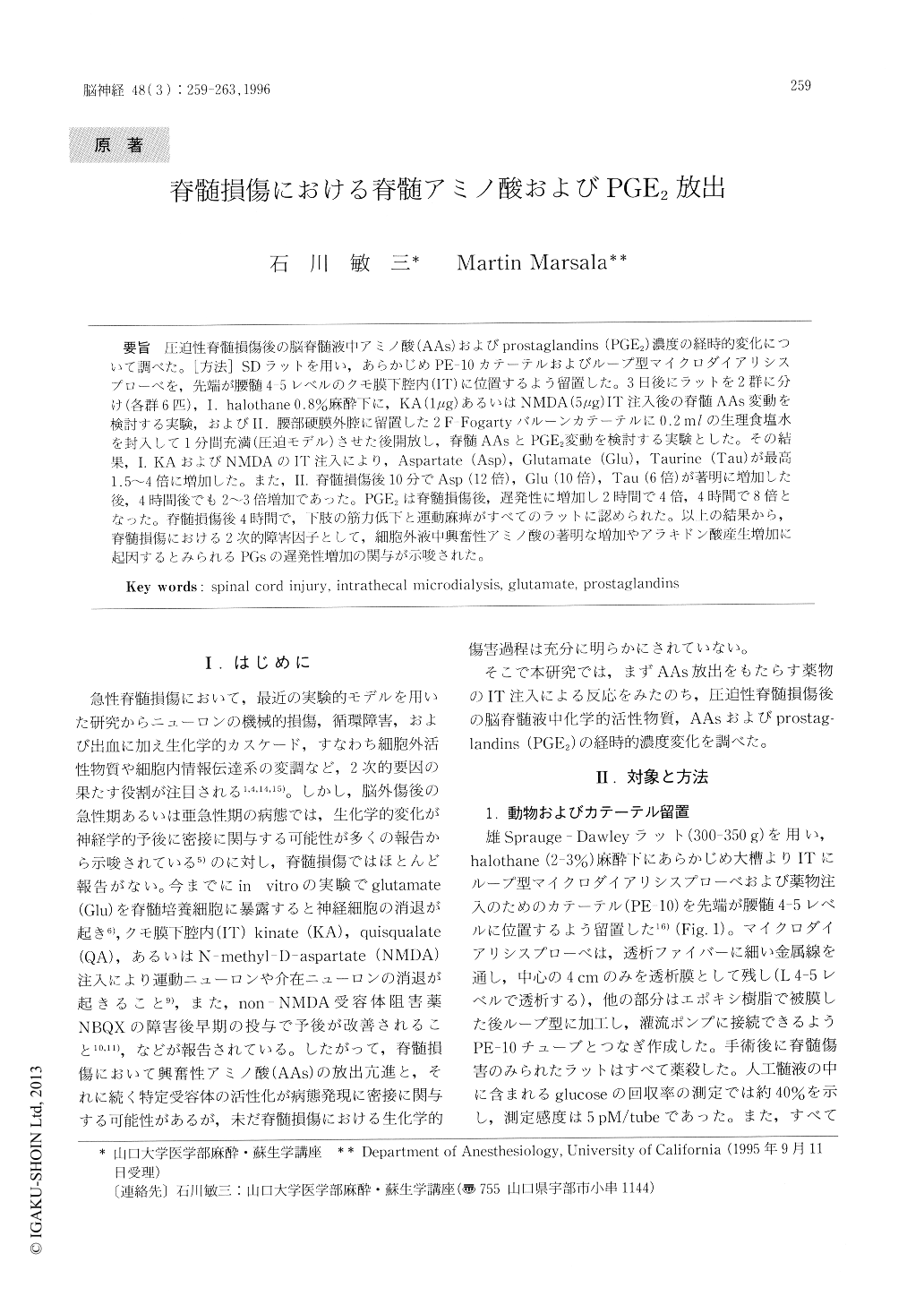
圧迫性脊髄損傷後の脳脊髄液中アミノ酸(AAs)およびprostaglandins(PGE2)濃度の経時的変化について調べた。[方法]SDラットを用い,あらかじめPE−10カテーテルおよびループ型マイクロダイアリシスプローベを,先端が腰髄4-5レベルのクモ膜下腔内(IT)に位置するよう留置した。3日後にラットを2群に分け(各群6匹),I.halothane 0.8%麻酔下に,KA(1μg)あるいはNMDA(5μg)IT注入後の脊髄AAs変動を検討する実験,およびII.腰部硬膜外腔に留置した2F-Fogartyバルーンカテーテルに0.2mlの生理食塩水を封入して1分間充満(圧迫モデル)させた後開放し,脊髄AAsとPGE2変動を検討する実験とした。その結果,1.KAおよびNMDAのIT注入により,Aspartate(Asp),Glutamate(Glu),Taurine(Tau)が最高1.5〜4倍に増加した。また,II.脊髄損傷後10分でAsp(12倍),Glu(10倍),Tau(6倍)が著明に増加した後,4時間後でも2〜3倍増加であった。PGE2は脊髄損傷後,遅発性に増加し2時間で4倍,4時間で8倍となった。脊髄損傷後4時間で,下肢の筋力低下と運動麻痺がすべてのラットに認められた。
Acute spinal cord trauma may initiate a cascade of hemodynamic and biochemical events characte-rized by direct mechanical destruction of neurons, hemorrhages and significant increases in active substances (glutamate, prostaglandins (PGs),ets.). However, there are no data which define the effect of acute spinal cord trauma on biochemical changes in the spinal cord. The aim of the present study was to examine time-dependent biochemical changes in the spinal cord by intrathecal microdialysis in the acute stage of spinal cord trauma.

Copyright © 1996, Igaku-Shoin Ltd. All rights reserved.


