Japanese
English
- 有料閲覧
- Abstract 文献概要
- 1ページ目 Look Inside
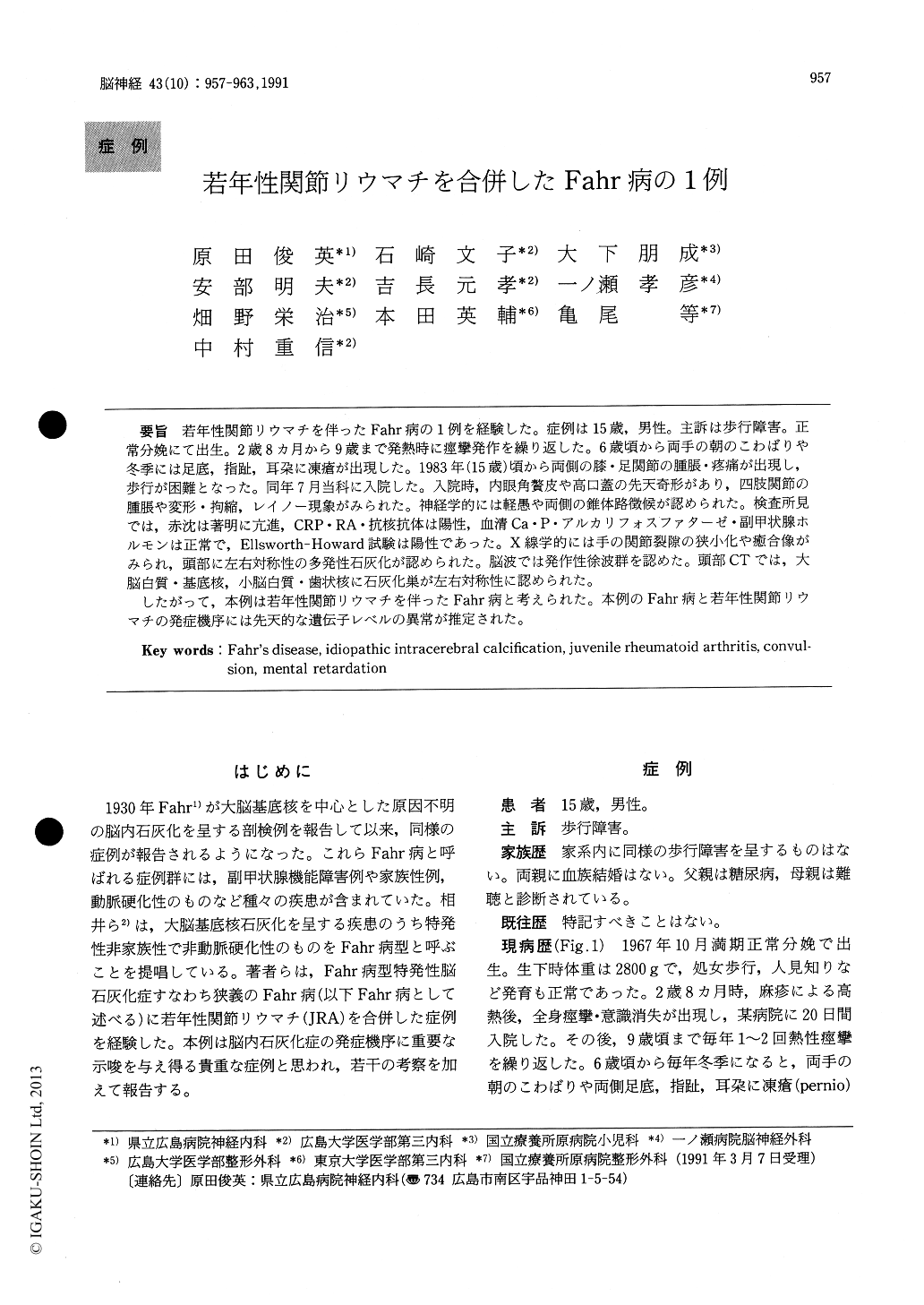
若年性関節リウマチを伴ったFahr病の1例を経験した。症例は15歳,男性。主訴は歩行障害。正常分娩にて出生。2歳8カ月から9歳まで発熱時に痙攣発作を繰り返した。6歳頃から両手の朝のこわばりや冬季には足底,指趾,耳朶に凍瘡が出現した。1983年(15歳)頃から両側の膝・足関節の腫脹・疼痛が出現し,歩行が困難となった。同年7月当科に入院した。入院時,内眼角贅皮や高口蓋の先天奇形があり,四肢関節の腫脹や変形・拘縮,レイノー現象がみられた。神経学的には軽愚や両側の錐体路徴候が認められた。検査所見では,赤沈は著明に亢進,CRP\RA\抗核抗体は陽性,血清Ca・P・アルカリフォスファターゼ・副甲状腺ホルモンは正常で,Ellsworth-Howard試験は陽性であった。X線学的には手の関節裂隙の狭小化や癒合像がみられ,頭部に左右対称性の多発性石灰化が認められた。脳波では発作性徐波群を認めた。頭部CTでは,大脳白質・基底核,小脳白質・歯状核に石灰化巣が左右対称性に認められた。
したがって,本例は若年性関節リウマチを伴ったFahr病と考えられた。本例のFahr病と若年性関節リウマチの発症機序には先天的な遺伝子レベルの異常が推定された。
We studied a case of Fahr's disease type idiopa-thic intracerebral calcification (Fahr's disease) as-sociated with juvenile rheumatoid arthiritis. The patient was a 15-year-old male with a chief com-plaint of gait disturbance. His family members had no similar signs and symptoms. His parents had no consanguinity. He was born with the normal perinatal course at 1967. He had repeated episodes of convulsive attacks during fever elevation from 2 yaers and 8 months to 9 years of age. Morning stiffness of bilateral hands, and pernio in the auri-cles, fingers, planta, and toes had occurred in everywinter, since 6 years old. Swelling and pain of the bilateral knee and foot joints appeared, making ambulation difficult in 1983 (15 years old), and the patient was admitted to our hospital in July, the same year.
On admission, congenital anomalies such as epicanthus and high-arched palate were noted, and swelling, deformation and contracture of limb joints, and Raynaud phenomenon were shown. His ocular fundus showed no arteriosclerotic change. He didn't have Albright's sign. Mild mental retarda-tion and bilateral pyramidal tract signs were noted, but extrapyramidal tract and cerebellar signs, and sensory disturbance were absent. Laboratory findings exhibited markedly elevated ESR, positive CRP, RA, and antinuclear antibody. The levels of serum Ca, P, alkaline phosphatase and parathyroid hormone were normal. Peripheral blood study showed microcytic and hypochromic anemia. Anti-DNA antibody was negative. Ellsworth-Howard test was positive. Elevated antibody titer to toxo-plasma, rubella virus, herpes simplex virus and cytomegalovirus were not proven. He had no chromosomal change. In the cerebrospinal fluid, the protein level was slightly high (49mg/dl), but the level of sugar, Na and Cl, and cell count were normal. Electromyographic examination had no abnormal change. Paroxysmal slow waves were shown in electroencephalogram. Roentogenogram revealed narrowing and fusions of the articular spaces in the hands, and symmetric multiple calcification in the head. Computed tomography confirmed symmetric calficication in the white matter and basal ganglia of the cerebrum, and the white matter and dentate nucleus of the cerebellum. Thus, the diagnosis of this patient was ruled out from the diseases, which might cause intracerebral calcifications, such as CO and lead intoxication, radiation, pseudohypoparathyroidism, pseudo-pseudohypoparathyroidism, toxoplasmosis, infec-tion of cytomegalovirus and rubella virus, tuberous sclerosis, Sturge-Weber disease, systemic lupus erythematodus, and Cockayne syndrome. He was considered to have Fahr's disease associated with juvenile rheumatoid arthritis. In this case, it is sug-gested that the pathogenesis of Fahr's disease and juvenile rheumatoid arthritis might be related with congenital genetic abnormalities.

Copyright © 1991, Igaku-Shoin Ltd. All rights reserved.


