Japanese
English
- 有料閲覧
- Abstract 文献概要
- 1ページ目 Look Inside
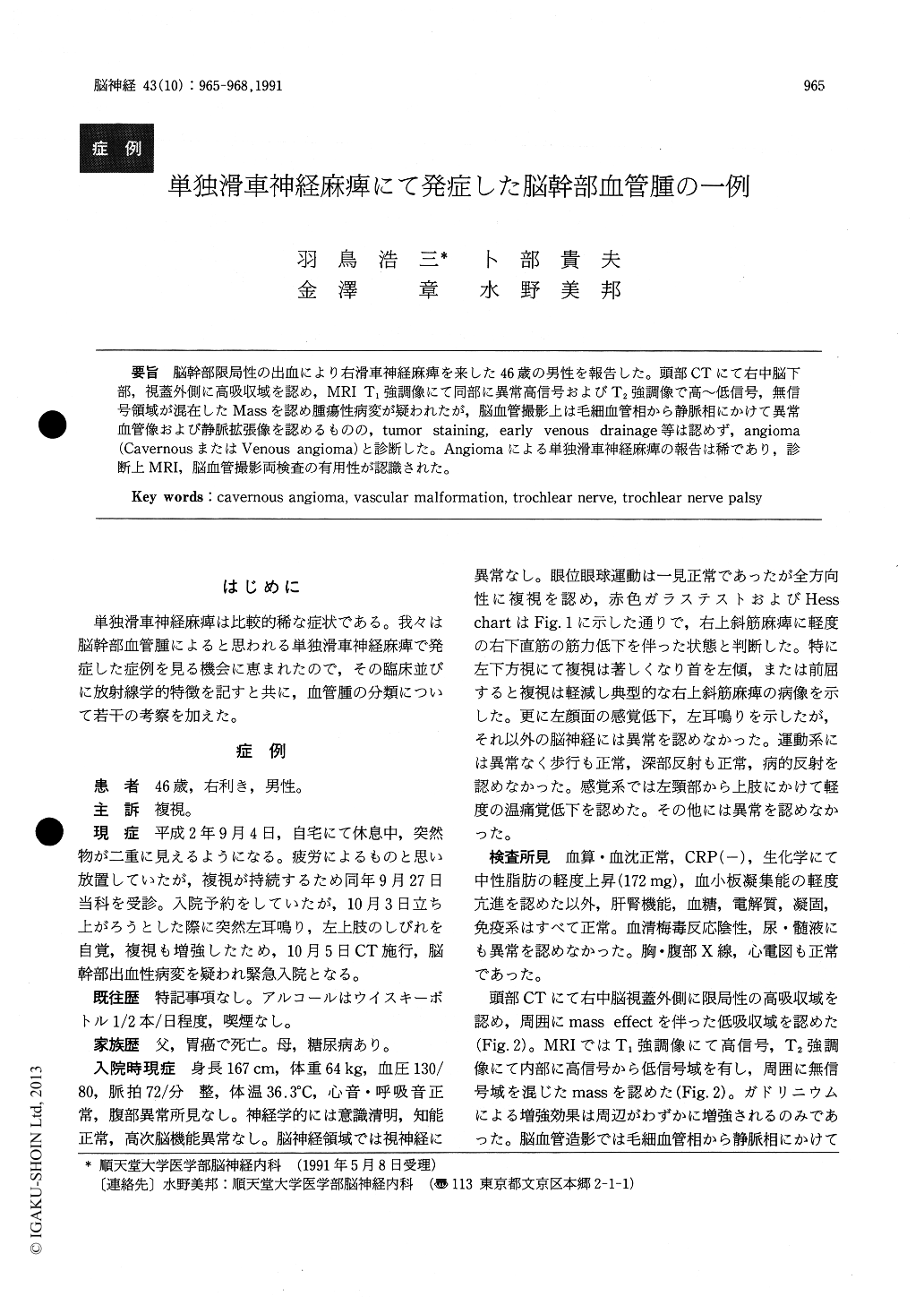
脳幹部限局性の出血により右滑車神経麻痺を来した46歳の男性を報告した。頭部CTにて右中脳下部,視蓋外側に高吸収域を認め,MRI T1強調像にて同部に異常高信号およびT2強調像で高〜低信号,無信号領域が混在したMassを認め腫瘍性病変が疑われたが,脳血管撮影上は毛細血管相から静脈相にかけて異常血管像および静脈拡張像を認めるものの,tumor stainingl early venous drainage等は認めず,angioma(CavernousまたはVenous angioma)と診断した。Angiomaによる単独滑車神経麻痺の報告は稀であり,診断上MRI,脳血管撮影両検査の有用性が認識された。
We report a 46-year-old, non-hypertensive man who suddenly developed isolated right trochlear nerve palsy. His diplopia was most prominent in the left lower gaze, and partially alleviated by head tilt to the left or by anteflexion of the neck. His CT scans showed a small high density area consistent with a hemorrhage in the lateral side of the right mesencephalic tectum.
His MRI (T2-weighted images) showed a lesion con-sisting of mixed high- and iso-intensity areas with linear low intensity areas. The margin of the lesion was irregular and nodular. Cerebral angiography (prolonged injection) showed small feeding arteries (or capellaries) in the late arterial phase and dilated draining veins in the venous phase. No tumor stain, early draining veins, or capillary brushes were present. We thought he had an angioma (vascular malformation). AVM seemed unlike-ly. Review of the literature revealed that trochlear nerve palsy caused by a mesencephalic angioma is extremely rare. MRI and cerebral angiography (prolonged injec-tion) seemed usefull for the diagnosis of angiomas (Vascular malformations).

Copyright © 1991, Igaku-Shoin Ltd. All rights reserved.


