Japanese
English
- 有料閲覧
- Abstract 文献概要
- 1ページ目 Look Inside
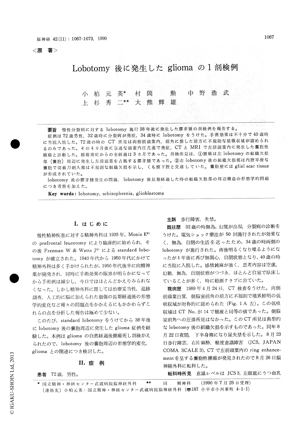
慢性分裂病に対するlobotomy施行38年後に発生した膠芽腫の剖検例を報告する。
症例は72歳男性,32歳時に分裂病が発症,34歳時にlobotomyをうけた。手術効果は不十分で40歳時に当院入院した。72歳の時のCT所見は両側前頭葉内,前角に接した前方に不規則な低吸収域が認められるのみであった。その4カ月後に急速な頭蓋内圧亢進で発症,CTとMRIで左前頭葉内に発生した嚢胞性腫瘍と診断した。腫瘍発症からの全経過は3カ月であった。剖検所見は,①腫瘍は左lobotomyの組織欠損部(嚢胞)周辺に発生し左前頭葉を占拠する膠芽腫であった。②右lobotomy後の組織欠損部は内腔平滑な嚢胞で切截刀刺入部は不規則な組織欠損を示し,くも膜下腔と交通していた。嚢胞壁にはglial scar tissueが形成されていた。
lobotomy後の膠芽腫発生の問題,lobotomy後長期経過した時の組織欠損部の周辺構造の形態学的問題につき考察を加えた。
The authors describe an autopsy case of glioblas-toma occurred after 38 years received lobotomy.
The patient was a 72 year-old male, who re-ceived lobotomy at 34 year old against schizo-phrenia. CT scan taken at 72 year old showed irregular low density areas without mass effect in the bilateral frontal white matter adjacent to the anterior horn.
After 4 months, the signs of intracranial hyper-tension were observed and his consciousness was disturbed abruptly. CT scan revealed ring en-hancement with marked mass effect in the left frontal lobe. A biopsy specimen from the tumor showed a picture of anaplastic astrocytoma. Family rejected the remission maintenance treatment. The patient died 3 months later the onset.
At autopsy, a large tumor occupied in the left frontal lobe was recognized. The tumor demarca-ted poorly from the cerebral tissue and invaded into the left anterior cingulate gyrus and the corpus callosum. Histologically, tumor cells com-posed of fibrillary, gemistocytic and multinucleatedastrocytes. GFAP, NSE and vimentin were found in large cells. Histological diagnisis was glioblas-toma. It was suggested that the tumor occurred from the region around a cyst of prefrontal lobo-tomy in the left frontal lobe.
In the right frontal lobe, a large cyst in size of 30 × 18 mm was present in the centrum semiovale. The wall of cyst was composed of layer of glial scar tissue. The origin of the cyst was discussed.

Copyright © 1990, Igaku-Shoin Ltd. All rights reserved.


