Japanese
English
- 有料閲覧
- Abstract 文献概要
- 1ページ目 Look Inside
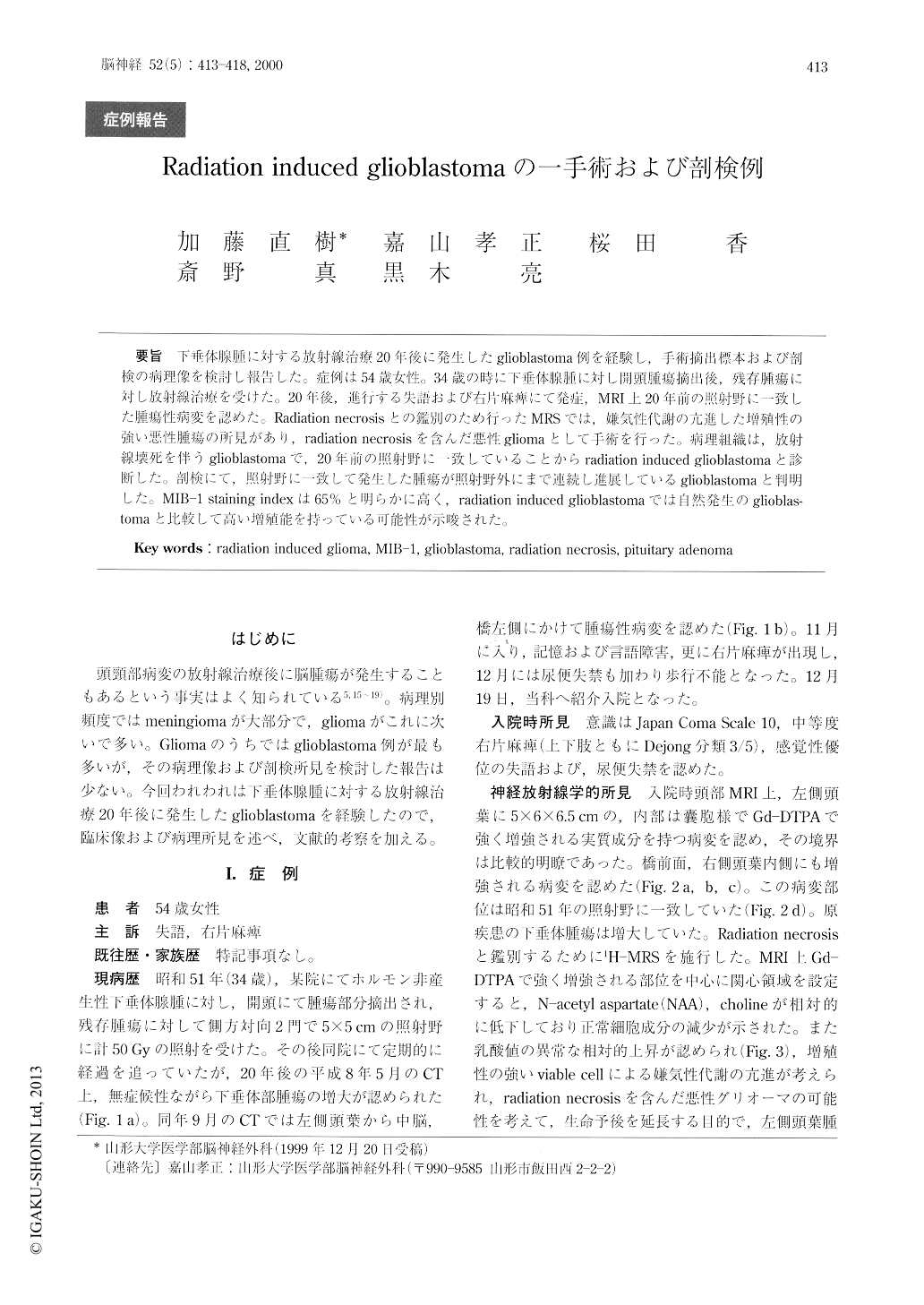
下垂体腺腫に対する放射線治療20年後に発生したglioblastoma例を経験し,手術摘出標本および剖検の病理像を検討し報告した。症例は54歳女性。34歳の時に下垂体腺腫に対し開頭腫瘍摘出後,残存腫瘍に対し放射線治療を受けた。20年後,進行する失語および右片麻痺にて発症,MRI上20年前の照射野に一致した腫瘍性病変を認めた。Radiation necrosisとの鑑別のため行ったMRSでは,嫌気性代謝の亢進した増殖性の強い悪性腫瘍の所見があり,radiation necrosisを含んだ悪性gliomaとして手術を行った。病理組織は,放射線壊死を伴うglioblastomaで,20年前の照射野に一致していることからradiation induced glioblastomaと診断した。剖検にて,照射野に一致して発生した腫瘍が照射野外にまで連続し進展しているglioblastomaと判明した。MIB-1 staining indexは65%と明らかに高く,radiation induced glioblastomaでは自然発生のglioblas—tomaと比較して高い増殖能を持っている可能性が示唆された。
We report a surgical case of a 54-year-old woman with a radiation induced glioblastoma. At the age of 34, the patient was diagnosed to have a non-function-ing pituitary adenoma. It was partially removed fol-lowed by 50 Gy focal irradiation with a 5×5 cm lateral opposed field. Twenty years later, she suffered from rapidly increasing symptoms such as aphasia and right hemiparesis. MRI showed a large mass lesion in the left temporal lobe as well as small mass lesions in the brain stem and the right medial temporal lobe. These lesions situated within the irradiated field.

Copyright © 2000, Igaku-Shoin Ltd. All rights reserved.


