Japanese
English
- 有料閲覧
- Abstract 文献概要
- 1ページ目 Look Inside
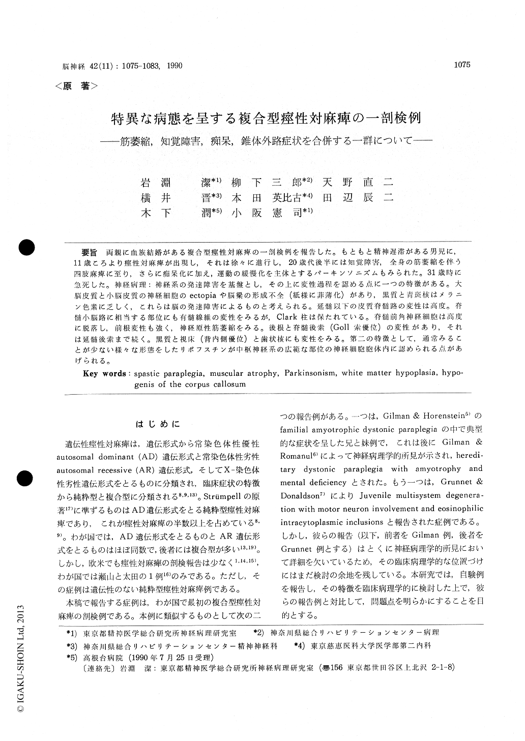
両親に血族結婚がある複合型痙性対麻痺の一剖検例を報告した。もともと精神遅滞がある男児に,11歳ころより痙性対麻痺が出現し,それは徐々に進行し,20歳代後半には知覚障害,全身の筋萎縮を伴う四肢麻痺に至り,さらに痴呆化に加え,運動の緩慢化を主体とするパーキンソニズムもみられた。31歳時に急死した。神経病理:神経系の発達障害を基盤とし,その上に変性過程を認める点に一つの特徴がある。大脳皮質と小脳皮質の神経細胞のectopiaや脳梁の形成不全(紙様に菲薄化)があり,黒質と青斑核はメラニン色素に乏しく,これらは脳の発達障害によるものと考えられる。延髄以下の皮質脊髄路の変性は高度。脊髄小脳路に相当する部位にも有髄線維の変性をみるが,Clark柱は保たれている。脊髄前角神経細胞は高度に脱落し,前根変性も強く,神経原性筋萎縮をみる。後根と脊髄後索(Goll索優位)の変性があり,それは延髄後索まで続く。黒質と視床(背内側優位)と歯状核にも変性をみる。第二の特徴として,通常みることが少ない様々な形態をしたリポフスチンが中枢神経系の広範な部位の神経細胞胞体内に認められる点があげられる。
An autopsied case of copmlicated form of spastic paraplegia with many unusual clinical and patho-logical features is reported.
Present case : a 31-year-old male. His parents are first cousins. Pregnancy and delivery had been unremarkable. Though he was mentally retarded, his physical development was normal. He was considered normal until age 10. He suf-fered from progressive disturbance in gait at the age of 11. He could not walk without assistance at the age of 22. Neurological examination re-vealed the following findings. He was obese and mentally deteriorated. Spastic paraplegia with increased tendon reflexes and pathological reflexes was prominent.
Though slight sensory disturbance was present in the lower extremities, neither involuntary move-ment nor cerebellar ataxia was observed. In the age of late 20's, dementia, general muscular atrophy, and Parkinsonism developed. At the age of 30, he could not move by himself. He was apathic and indifferent, and showed forced laughing. Muscle tonus was flaccid because of general muscular atrophy and peripheral neuro-pathy. He died of acute gastric enlargment.
Neuropathological findings were characterized by mal-development of the central nervous system (CNS) and the multisystem degeneration. There exisited cerebral white matter hypoplasia with hypogenesis of the corpus callosum and ectopia of neurons of the cerebral and cerebellar cortex. Hypoplasia of melanin pigment was also observed in the remaining neurons of the substantia nigra and the locus ceruleus. Many neurons in the CNS included lipofuscin granules of variable shapes. Some of them showed clusters of several block-like inclusions which were green with luxol fast blue and cresyl violet stain. In addition, re-markable degeneration in the pyramid of the medulla oblongata and the lateral and anterior cortico-spinal tracts of the spinal cord was ob-served. The dosal colum showed myelin pallor predominent in the Goll's fasiculus. There was moderate neuronal loss in the spinal anterior horn and dorsal root ganglia. Degeneration was much more severe in the anterior roots than in the posterior roots of the spinal nerves. Skeletal muscles revealed marked group atrophy. There were moderate neuronal loss in the substantia nigra and the dorso-medial nucleus, pulvinar, and lateral geniculate body of the thalamus, slightneuronal loss with grumose degeneration in the dentate nucleus, and fibrous gliosis in the vestibu-lar nuclei. Neuropathological and biochemical studies could not reveal certain metabolic diseases.
Clinico-pathological findings of our case resem-ble those of the cases reported by Gilman et al (1964, 1975) and Grunnet & Donaldson (1985). The parents of these families including of our family are first cousins. In conclusion, we propose that this kind of complicated form of hereditary spast-ic paraplegia is a clinico-pathological entity.

Copyright © 1990, Igaku-Shoin Ltd. All rights reserved.


