Japanese
English
- 有料閲覧
- Abstract 文献概要
- 1ページ目 Look Inside
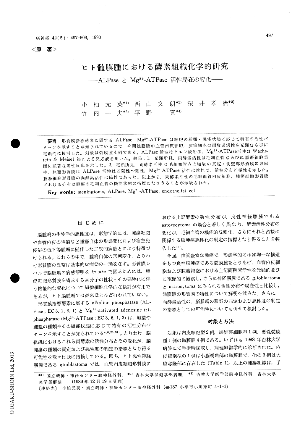
形質膜指標酵素に属するALPase,Mg2+-ATPaseは細胞の種類.機能状態に応じて特有の活性パターンを示すことが知られているので,今回髄膜腫の血管内皮細胞,腫瘍細胞の両酵素活性を光顕なりびに電顕的に検討した.対象は髄膜腫4例である。ALPase活性はクエン酸鉛法,Mg2+-ATPase活性はWachstein & Meisel法による反応液を用いた。結果:1.光顕所見,両酵素活性は毛細血管ならびに腫瘍細胞集団に顕著な陽性反応を示した。2.電顕所見,両酵素活性は毛細血管内皮細胞の基底・側壁部形質膜に強陽性,腔面形質膜はALPase活性は弱陽性〜陰性,Mg2+-ATPase活性は陰性で,活性分布に極性を示した。腫瘍細胞形質膜の両酵素活性は陽性であった。以上から,両酵素活性の毛細血管内皮細胞,腫瘍細胞形質膜における分布は腫瘍の毛細血管の機能状態の指標になりうることが示唆された。
Ultracytochemical features of microvessels and tumor cells of the human meningiomas were examined by light and electron microscopy with special reference to the distribution of Mg2+-ATPase and alkaline phosphatase (ALPase) activity on the walls of the vessels and tumor cell surfaces.
Materials used were 4 cases of meningiomas, 2 of which were meningotheliomatous type, one fibroblastic type and one malignant meningioma respectively.
For ultracytochemistry, specimens were quickly fixed in an ice-cold 0.1 M cacodylate buffer containing 8% sucrose (pH 7. 2) for one hour and transferred to a substrate solution for detection of Mg2+-ATPase and ALPase. The preparations were incubated at 37℃ for 15-30 min in the medium described by Mayahara et al. for ALPase and for 15-20 min in the medium described by Wachstein and Meisel. The control samples were incubated in a medium containing 1 mm Bromotetramisole for ALPase and also in a substrate free medium for Mg2+-ATPase.
At the light microscopy, Mg2+-ATPase and AL-Pase activities appeared to be mainly restricted to the capillary wall and around or in the tumor cell nest showing whorl formation. Both enzyme activities were negative in the control study.
By electron microscopy, reaction products representing Mg2+-ATPase activity were distributed in the basolateral plasma membrane of the endothelial cells on the surface of the pericytes and on the surface of the tumor cells. Reaction products of ALPase activity located mainly on the abluminal surfaces of the endothelial cells and in some specimen on both luminal and abluminal surfaces ofthose cells. Intense reaction products were distributed evenly on all round surfaces of the tumor cells.
Changes in distribution pattern of ALPase activity such as on the abluminal surfaces in low grade cases and on the luminal one in high grade cases were reported in human glioma vessels. In this study, these enzyme activities were positive not only on the endothelial cells, but on the surfaces of the human meningioma cells.
The reaction products of the endothelial cell membrane enzymes were not evenly distributed between the luminal and abluminal plasma membrane. Thus, the degree of changes in the distribution of enzyme activities was different for each enzyme, grade of tumor and nature of tumor. These regional differences in these enzyme cytochemistry may represent functional heterogeneity in the endothelial cell membrane of the human brain tumors.

Copyright © 1990, Igaku-Shoin Ltd. All rights reserved.


