Japanese
English
- 有料閲覧
- Abstract 文献概要
- 1ページ目 Look Inside
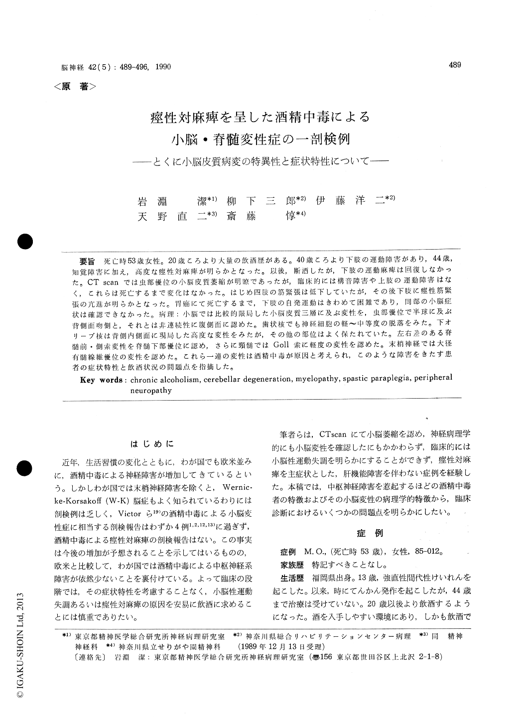
死亡時53歳女性。20歳ころより大量の飲酒歴がある。40歳ころより下肢の運動障害があり,44歳,知覚障害に加え,高度な痙性対麻痺が明らかとなった。以後,断酒したが,下肢の運動麻痺は回復しなかった。CT scanでは虫部優位の小脳皮質萎縮が明瞭であったが,臨床的には構音障害や上肢の運動障害はなく,これらは死亡するまで変化はなかった。はじめ四肢の筋緊張は低下していたが,その後下肢に痙性筋緊張の充進が明らかとなった。胃癌にて死亡するまで,下肢の自発運動はきわめて困難であり,同部の小脳症状は確認できなかった。病理:小脳では比較的限局した小脳皮質三層に及ぶ変性を,虫部優位で半球に及ぶ背側面吻側と,それとは非連続性に腹側面に認めた。歯状核でも伸経細胞の軽〜中等度の脱落をみた。下オリーブ核は背側内側面に現局した高度な変性をみたが,その他の部位はよく保たれていた。左右差のある脊髄前・側索変性を脊髄下部優位に認め,さらに頸髄ではGoll索に軽度の変性を認めた。末梢神経では大径有髄線維優位の変性を認めた。これら一連の変性は酒精中毒が原因と考えられ,このような障害をきたす患者の症状特性と飲酒状況の問題点を指摘した。
A 45 year-old-female was admitted to Kanagawa Rehabilitation Center because of marked spastic paraplegia. There was no family history of neu-rological diseases. She had been drinking a great excess of alcohol since twenty years of age. Tho-ugh she noticed the unsteadiness of gait several years prior to the admission, she had not been exa-mined.
On admission neurological examination revealed pyramidal weakness of both legs, and slight sen-sory impairment in distal part of the lower extre-mities. But she had neither difficulty in speech nor abnormal findings in the upper extremities. Her mentality was well preserved. Though com-puted tomography revealed cerebellar cortical at-rophy, no signs of cerebellar impairment could not be found except spastic paraplegia. Blood chemi-stry failed to reveal liver dysfunction. There was no pernicious anemia. These symptoms were al-most stationary until her death. At the age of 53, she died of gastric cancer.
Postmortem examination disclosed marked dege-neration of cerebellum and spinal cord. Cerebel-lar cortical degeneration, which was characterized by involvement of all layers of the cortex, showed unique distribution. The lingula, central lobule, culumen, superior portion of declive, anterior lobu-les and anteromedial half of simple lobule wereseverely degenerated, while other lobules were spared excluding moderate degeneration in infero-medial portion of the hemisphere. These cortical degeneration was prominent much more in vermis than in hemisphere. There was a mild myelin pal-lor in the lamellar white matter and slight fibrous gliosis. In the dentate nucleus, there were mode-rate neuronal loss with gliosis. In the inferior oli-vary complexes, there was a complete loss of neu-rons with accompanying gliosis in the dorso-medial portion and cap of the dorsal lamina and dorsal accessary olives while neurons in the lateral and ventral portions were spared. Myelin pallor ex-sited in the lateral and anterior colums of the spi-nal cord, especially prominent in the lower seg-ments of the spinal cord. Slight myelin pallor was found in Goll's fasciculus of the cervical spinal cord. There were slight neuronal loss in the an-terior horn of the spinal cord and dorsal ganglion. As to the peripheral nerves, marked degeneration was obvious mostly in large myelinated fibers.
These characteristic degeneration of cerebellar cortex was identical with alcoholic cerebellar de-generation which was established by Victor et al (1959), although clinical examination failed to re-veal any signs of cerebellar involvement because of severe spastic paraplegia. Alcoholic cerebellar degeneration is charaterized by unique clinical sy-mptoms ; cerebellar ataxia is affected much more in the lower extremities than in the upper extre-mities, and speech is spared. Therefore, we did not observe any cerebellar symptoms, because she could not move her legs voluntarily because of severe pyramidal involvement. Recently, alcoho-lic spastic paraplegia without liver dysfunction has been drawing attention but there has been no neu-ropathological study for the present. These pa-thological changes should be considered to be cau-sed by chronic alcoholic intoxication ; cerebellar degeneration, myelopathy, and peripheral neuro-pathy.

Copyright © 1990, Igaku-Shoin Ltd. All rights reserved.


