Japanese
English
- 有料閲覧
- Abstract 文献概要
- 1ページ目 Look Inside
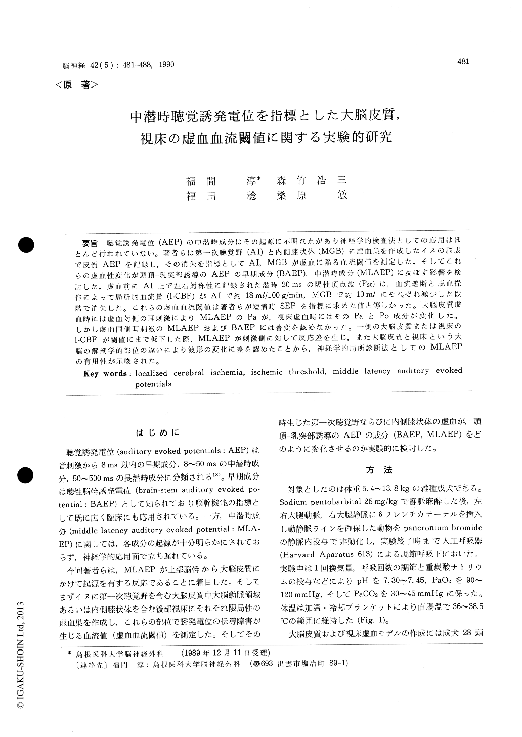
聴覚誘発電位(AEP)の中潜時成分はその起源に不明な点があり神経学的検査法としての応用はほとんど行われていない。著者らは第一次聴覚野(AI)と内側膝状体(MGB)に虚血巣を作成したイヌの脳表で皮質AEPを記録し,その消失を指標としてAI, MGBが虚血に陥る血流閾値を測定した。そしてこれらの虚血性変化が頭頂—乳突部誘導のAEPの早期成分(BAEP),中潜時成分(MLAEP)に及ぼす影響を検討した。虚血前にAI上で左右対称性に記録された潜時20msの陽性頂点波(P20)は,血流遮断と脱血操作によって局所脳血流量(l-CBF)がAIで約18ml/100g/min, MGBで約10mlにそれぞれ減少した段階で消失した。これらの虚血血流閾値は著者らが短潜時SEPを指標に求めた値と等しかった。大脳皮質虚血時には虚血対側の耳刺激によりMLAEPのPaが,視床虚血時にはそのPaとPo成分が変化した。しかし虚血同側耳刺激のMLAEPおよびBAEPには著変を認めなかった。一側の大脳皮質または視床のl-CBFが閾値にまで低下した際,MLAEPが刺激側に対して反応差を生じ,また大脳皮質と視床という大脳の解剖学的部位の違いにより波形の変化に差を認めたことから,神経学的局所診断法としてのMLAEPの有用性が示唆された。
Middle latency auditory evoked potentials (ML-AEPs) have not yet been established as a device of assessing neurological events because their origins have not been definitely identified. In this study, we assessed the neurological usefulness of MLA-EPs through experimental approach using canine models of acute ischemia localized within the cere-bral cortex or thalamus.
Two types of localized cerebral ischemia were produced in mongrel dogs by clipping of major ce-rebral arteries and inducing of hypotension ; they were, unilateral cortical ischemia involving the right primary auditory area (group A) and unila-teral thalamic ischemia involving the right medial geniculate body (group B). Using these two ische-mia models, auditory evoked potentials (AEPs) were intracranially and extracranially recorded in addition to the measurement of local cerebral blo-od flow (1-CBF).
Prior to the induction of ischemia, it was confir-med that positive waves with a latency of 20 ms (P20) were evoked in the bilateral primary auditory areas by sound stimulation (90 dB 5 Hz click) given to the ear contralateral to the planned ischemic side in both groups. The right P20 disappeared when the 1-CBF in the right primary auditory area decreased below the ischemic flow threshold of synaptic transmission failure, approximately 18 ml/ 100 g/min, in group A, and when the 1-CBF in the right medial geniculate body decreased below the threshold, 10 ml/100 g/min, in group B, These is-chemic thresholds determined by using a loss of cortical AEP response as an indicator in the pre-sent study showed no statistical differences from those determined by using a loss of cortical resp-onse with somatosensory evoked potential (SEP) as an indicator in the same canine models in our pre-vious study. After the right P20 disappeared, far-field potentials remained in both groups. These residual waves, which were thought to probably originate from lower generators, such as medial geniculate body, inferior colliculus and lateral le-mniscus, showed the highest positive peaks with a latency of 9. 2 ms in group A and that of 4. 5 ms in group B. In group A, a depth electrode placed in the medial geniculate body recorded the highest positive potential with the same latency as that preceding the highest positive residual potential, suggesting that the positive residual wave with a latency of 9. 2 ms was generated from the medial geniculate body. Residual potentials observed in group B were thought to be brain-stem auditory evoked potentials (BAEPs) recorded by a cortical surface electrode.
In regard to extracranially-recorded AEPs, BA-EPs or MLAEPs by ipsilateral ear (ischemic side) stimulation showed no remarkable changes in ei-ther group in comparison with those recorded be-fore the induction of ischemia. On the other hand, MLAEPs by contralateral ear (non-ischemic side) stimulation altered when a 1-CBF decreased below an ischemic threshold ; namely, Pa disappeared in both groups, and Po also decreased in amplitude in group B.
To summarize these results, changes in MLAEP waveforms differ between cortical ischemia and thalamic ischemia ; when 1-CBF in unilateral cere-bral cortex or thalamus decreased below an ische-mic threshold, the response to contralateral ear sti-mulation differed to that to ipsilateral ear stimula-tion. Thus we thought that MLAEPs are useful for the assessment of functional changes caused by unilateral lesion (s) in the posterior thalamus and/or temporal lobe.

Copyright © 1990, Igaku-Shoin Ltd. All rights reserved.


