Japanese
English
- 有料閲覧
- Abstract 文献概要
- 1ページ目 Look Inside
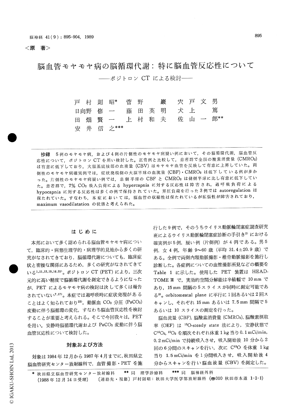
抄録 5例のモヤモヤ病,および4例の片側性のモヤモヤ病疑い例において,その脳循環代謝,脳血管反応性について,ポジトロンCTを用い検討した。正常例と比較して,患者群で全脳の酸素消費量(CMRO2)は有意に低下しており,大脳基底核部の血液量(CBV)はモヤモヤ血管を反映して有意に上昇していた。両側性のモヤモヤ病確実例では,症状発現側の大脳半球の血流量(CBF)・CMRO2は低下している例が多かった。片側性のモヤモヤ病疑い例では,患側半球のCBFとCMRO2は健側半球に比し有意に低下していた。患者群で,7%CO2吸入負荷によるhypercapniaに対する反応性は障害され,過呼吸負荷によるhypercapniaに対する反応性は多くの例で保持されていた。昇圧負荷を行った3例ではautoregulationは保たれていた。すなわち,本症においては,脳血管の収縮性は保たれているが拡張性が障害されており,maximum vasodilatationの状態と考えられた。
We investigated cerebral blood flow and meta-bolism, and cerebral vascular response in 9 patients with cerebrovascular Moyamoya disease or unilate-ral Moyamoya phenomenon using positron emis-sion tomography (PET). The subjects consisted of 5 men and 4 women, and were from 9 to 60 years old. Five patients had bilateral occlusion in the carotid fork with Moyamoya vessels (fulfiled the criteria of cerebrovascular Moyamoya disease), and four patients had unilateral Moyamoya phe-nomenon. The PET scanner used was the HEAD-TOME III, of which spatial resolution in clinical use was 10mm full width at half-maximum (FWHM) in the image plane. Cerebral blood flow (CBF), cerebral metabolic rate of oxygen (CMRO2), cere-bral oxygen extraction fraction (OEF), and cerebral blood volume (CBV) were measured in resting state by the 15O-labelled gases steady state method in every patient and 22 normal controls (17 men and 5 women, and from 26 to 64 years old). Con-secutively cerebral vascular responses were mea-sured by H215O autoradiographic method in resting state, hypercapnia, hypocapnia, and hypertension. Forced hypercapnia, hypocapnia, and hypertension were achieved by 7% CO2 inhalation, hyperventi-lation, and venous infusion of angiotensin I[, re-spectively.
CMRO2 of the whole brain was significantly lower in patients than in normal controls (p<0. 05), and CBV of the lentiform nucleus significantly increased in patients (p<0. 01). This reflected Moyamoya vessels in the basal ganglionic regions. In 3 of 5 patients with bilateral Moyamoya ves-sels, CBF and CMRO2 in the symptomatic cere-bral hemisphere were lower than that in the nonsymptomatic hemisphere. In all of 4 patients with unilateral Moyamoya phenomenon, CBF and CMROz of cerebral hemisphere on the side of the lesion were lower than those on the other side.
Vascular response in hypercapnia was impairedin patients compared with that in normal controls (p<0.05), while vascular response in hypocapnia and hypertension was maintained in most patients. Vascular response of the cerebral hemisphere on the side of Moyamoya vessels in patients with uni-lateral Moyamoya phenomenon was impaired in hypercapnia and hypocapnia compared with that of the cerebral hemisphere on the contralateral side of Moyamoya vessels. These indicate im-paired vasodilative response, and maintained vaso-contractive response, suggesting maximal dilata-tion of peripheral vessels in patients with Moya-moya disease. The maintained vasocontractivity may coincide with the symptom which is apt to appear in hyperventilation such as crying. How-ever, vascular response in hypocapnia was im-paired in one patient with bilateral Moyamoya vessels who showed marked impairment of vaso-reactivity for hypercapnia.

Copyright © 1989, Igaku-Shoin Ltd. All rights reserved.


