Japanese
English
- 有料閲覧
- Abstract 文献概要
- 1ページ目 Look Inside
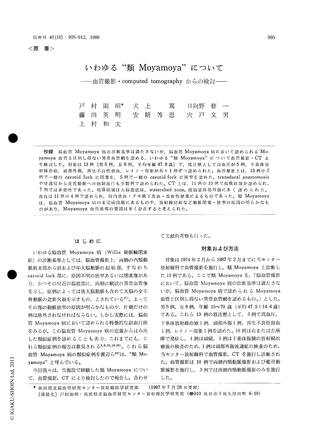
抄録 脳血管Moyamoya病の診断基準は満たさないが,脳血管Moyamoya病において認められるMo-yamoya血管と区別し得ない異常血管網を認める,いわゆる"類Moyamoya"について血管撮影・CTより検討した。対象は13例(男5例,女8例,平均年齢47.9歳)で,既往歴として高血圧が5例,下垂体放射線治療,頭部外傷,再生不良性貧血,レイノー現象が各々1例ずつ認められた。血管撮影上は,13例中7例で一側のcarotid forkに閉塞を,5例で一側のcarotid forkに狭窄を認めた。transdural anastomosisや穿通枝から皮質動脈への側副血行も少数例で認められた。CT上は,11例中10例で低吸収域が認められ,7例では多発性であった。低吸収域は大脳基底核,watershed zone,側脳室体部外側に多く認められた。出血は11例中4例で認められ,脳内出血・クモ膜下出血・出血性梗塞によるものであった。類Moyamoyaは,脳血管Moyamoya病の未完成段階にあるものや,放射線照射など動脈閉塞・狭窄の原因の明らかなものがあり,Moyamoya血管出現の要因は多く存在すると考えられた。
The criteria of the cerebrovascular Moyamoya disease is defined by the characteristic findings of its cerebral angiograms, as follows ; 1) The internal carotid siphon is narrowed or obstructed bilate-rally. 2) The "Moyamoya vessels" are observed at the base of the brain or the basal ganglionic regions. 3) Main trunks of the cerebral arteries such as the anterior, the middle, and/or the posteri-or cerebral arteries are often not or poorly visu-alized. 4) Its etiology is unknown. It has been known that the occlusion of the internal carotid fork with Moyamoya vessels is not infrequently seen in patients with tuberculous meningitis, sickle cell anemia, head trauma, and so on. In the defi-nition of the disease, patients with known etiology and/or unilateral occlusion in the carotid fork mustbe excluded. However, the cases who cannot fulfil its criteria of the cerebrovascular Moyamoya disease, but have its characteristic Moyamoya ves-sels and collateral pathways have been reported.
We investigated the findings of cerebral compu-ted tomograms in 13 patients who did not fulfil the criteria of the cerebrovascular Moyamoya dis-ease, but revealed the Moyamoya vessels. The sub-jects are 5 males and 8 females, ranging 15 to 70 years old. The past histories of 9 patients among them revealed hypertension, radiation therapy for pituitary adenoma, head trauma, aplastic anemia, and the Rainoud phenomenon. By angiographic evaluations, occlusions in the unilateral carotid forks were seen in 7 patients, and stenoses in those were in 5 patients. One patient showed only a severe stenosis in the horizontal portion of the middle cerebral artery. Twelve patients showed the Moyamoya vessels at the base of the brain called "basal Moyamoya", and 2 patients showed the vessels in the cranial vault called "vault Mo-yamoya", and a patient showed this abnormality in the ethmoid sinus and nasal cavity called "ethmoid Moyamoya". Transdural anastomosis via the superficial temporal arteries, the anterior ethmoid arteries, etc., and the collateral pathway from the perforating branches to the cortical branches were seen in a few patients. It was con-sidered that the collateral pathway from the per-forating branches to the cortical branches was newly developed flow via the ventriculofugal me-dullary arteries ; transmedullary anastomosis.
Computed tomograms revealed multiple low density areas in 10 of 11 patients. The location of low density area on CT scan was mostly in the basal ganglionic regions which was rare in genu-ine Moyamoya-disease, watershed zones, and lateral to the body of the lateral ventricles. Four pa-tients showed intracranial hemorrhage, such as intracerebral hematoma, subarachnoid hemorrhage due to ruptured aneurysm, and hemorrhagic infarc-tion.
The present subjects were similar to the cere-brovascular Moyamoya disease not only in the neuroradiological findings but also in the clinical symptoms. It was considered that the patients with known etiology such as radiation therapy, aplastic anemia, and head trauma could be differen-tiated from patients with the cerebrovascular Moyamoya disease, but patients with unknown etiology might be incomplete form of the cerebro-vascular Moyamoya disease.

Copyright © 1988, Igaku-Shoin Ltd. All rights reserved.


