Japanese
English
- 有料閲覧
- Abstract 文献概要
- 1ページ目 Look Inside
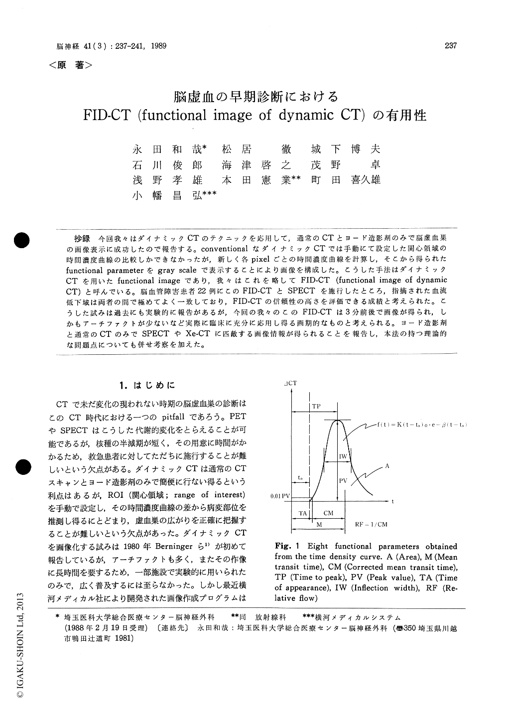
抄録 今回我々はダイナミックCTのテクニックを応用して,通常のCTとヨード造影剤のみで脳虚血巣の画像表示に成功したので報告する。conventionalなダイナミックCTでは手動にて設定した関心領域の時間濃皮曲線の比較しかできなかったが,新しく各pixelごとの時間濃度曲線を計算し,そこから得られたfunctional parameterをgray scaleで表示することにより画像を構成した。こうした手法はダイナミックCTを用いたfunctional imageであり,我々はこれを略してFID-CT (functional image of dynamicCT)と呼んでいる。脳血管障害患者22例にこのFID-CTとSPECTを施行したところ,指摘された血流低下域は両者の間で極めてよく一致しており,FID-CTの信頼性の高さを評価できる成績と考えられた。こうした試みは過去にも実験的に報告があるが,今回の我々のこのFID-CTは3分前後で画像が得られ,しかもアーチファクトが少ないなど実際に臨床に充分に応用し得る画期的なものと考えられる。ヨード造影剤と通常のCTのみでSPECTやXe-CTに匹敵する画像情報が得られることを報告し,本法の持つ理論的な問題点についても併せ考察を加えた。
A computer program was newly developed to display the ischemic area from the serial dynamic CT scan, namely the functional image of the dynamic CT (FID-CT). The principles of FID-CT are as follows ; Seven rapid-sequence dynamic CT scans were taken following a peripheral bolus intravenous injection of 40 ml of iopamidol. As the data from each scan can be separated into three consecutive segments, we obtained 21 images during 44 seconds. Time density curves of each pixel were calculated employing Gamma variate fitting method. Eight functional parameters obtain-ed from this curve were displayed with gray-scale pixel by pixel. These eight images were functional images obtained from dynamic CT. Only three or four minutes were required to complete all the calculations.
Twenty-two patients were examined with both FID-CT and 123I-SPECT. In each case, the lesion detected by FID-CT was remarkably consistent with that shown by SPECT. Two representative cases were presented. The authors believe that FID-CT is a very useful diagnostic method in the acute stage of cerebral ischemia because the method can be quickly and easily performed and it discloses the ischemic area with fair certainty.

Copyright © 1989, Igaku-Shoin Ltd. All rights reserved.


