Japanese
English
- 有料閲覧
- Abstract 文献概要
- 1ページ目 Look Inside
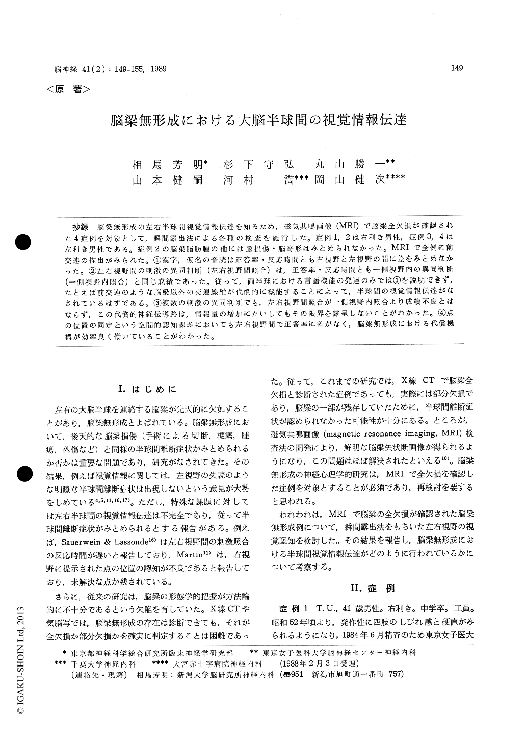
抄録 脳梁無形成の左右半球間視覚情報伝達を知るため,磁気共鳴画像(MRI)で脳梁全欠損が確認された4症例を対象として,瞬間露出法による各種の検査を施行した。症例1, 2は右利き男性,症例3, 4は左利き男性である。症例2の脳梁脂肪腫の他には脳損傷・脳奇形はみとめられなかった。MRIで全例に前交連の描出がみられた。①漢字,仮名の音読は正答率・反応時間とも右視野と左視野の間に差をみとあなかった。②左右視野間の刺激の異同判断(左右視野間照合)は,正答率・反応時間とも一側視野内の異同判断(一側視野内照合)と同じ成績であった。従って,両半球における言語機能の発達のみでは①を説明できず,たとえば前交連のような脳梁以外の交連線維が代償的に機能することによって,半球間の視覚情報伝達がなされているはずである。③複数の刺激の異同判断でも,左右視野間照合が一側視野内照合より成績不良とはならず,この代償的神経伝導路は,情報量の増加にたいしてもその限界を露呈しないことがわかった。④点の位置の同定という空間的認知課題においても左右視野間で正答率に差がなく,脳梁無形成における代償機構が効率良く働いていることがわかった。
It has been well established that acquired lesions of the corpus callosum such as surgical section bring about disturbances of interhemi-spheric transfor of visual information. In contrast, patients with callosal agenesis do not display these specific deficits. The mechanisms of this compensation have been postulated as follows;
(1)bilateral development of language function,
(2) exploitation of extracallosal commissure fibers such as the anterior commissure.
Several studies have reported, however, minor disturbances of interhemispheric visual transfer in callosal agenesis, such as less efficient inter-hemispheric transfer of complex visual stimuli (Gott & Saul, 1978), slower reaction time in interhemispheric comparison of visual stimuli (Sauerwein & Lassonde, 1983), or deficits in spatial localization in the right hemi-field (Martin, 1985)
In order to settle these issues, we have admi-nistered four kinds of tachistoscopic visual recognition tests on 4 patients with complete agenesis of the corpus callosum confirmed by magnetic resonance imaging (MRI). This technique enabled us to see the mid-sagittal plane of the corpus callosum and diagnose its total absence with much higher certainty and precision than previous studies employing computed tomography (CT) or pneumoencephalography.
Case 1 : A 41-year-old right-handed man visited us because of recurrent numbness in the four extremities. Neurological examination revealedno abnormalities. MRI has confirmed that the corpus callosum was totally lacking, and that the anterior commissure was normally visualized.
Case 2: A 31-year-old right-handed man was referred to us for the treatment of partial complex seizure. Total agenesis and lipoma of the corpus callosum was diagnosed by MRI and reconstructed sagittal view of CT scan. MRI in-dicated the existence of the anterior commissure.
Case 3: A 24-year-old left-handed man was admitted to our hospital under the diagnosis of aseptic meningitis. Except for the median cleft face syndrome, neurological examination was normal when he recovered from meningitis. MRI has revealed total callosal agenesis and the an-terior commissure.
Case 4: A 32-year-old left-handed man was admitted to our hospital under the diagnosis of amyotrophic lateral sclerosis. MRI revealed total callosal agenesis and the normal anterior corn-missure.
Test 1 : Reading aloud of unilaterally presented letters (Kanji & Kana). All the patients correctly read aloud all of the stimuli, showing that unilateral alexia did not exist. Reaction time of reading aloud did not differ between right and left visual fields.
Test 2: Inter-visual field and intra-visual field same/different comparison tasks of verbal stimuli were compared. The difference was not signifi-cant either in percentage of correct response or reaction time.
The result of Test 2 suggests that exploitation of some kind of extracallosal commissure fibers must take place in callosal agenesis. Judging from the equal reaction time this compensation system seemed to be quite efficient.
Test 3: Inter-visaul field and intra-visual field matching of pleural stimuli were also equally efficient, which suggests that the extracallosal fiber pathway does not expose its limit of functional compensation even when it is loaded with heavier information transfer.
Test 4: Dot localization task was equally per-formed in either visual field. It is suggested that this sort of spatial information also transfers efficiently through the extracallosal compen-satory pathway.

Copyright © 1989, Igaku-Shoin Ltd. All rights reserved.


