Japanese
English
- 有料閲覧
- Abstract 文献概要
- 1ページ目 Look Inside
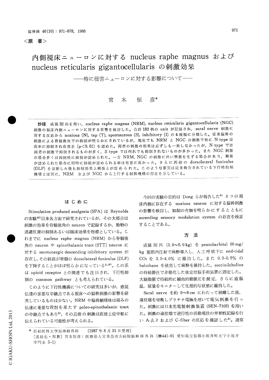
抄録 成猫32匹を用い,nucleus raphe magnus (NRM), nucleus reticularis gigantocellularis (NGC)刺激の視床内側ニューロンに対する影響を検討した。合計183個のunitが記録され,sural nerve刺激に対する反応からnoxious (N),tap (T), spontaneous (S), inhibitory (I)の4種類に分類した。従来脳幹の刺激による脊髄後角での抑制が明らかにされているが,視床でもNRMとNGCの刺激で特にN typeは高率に抑制され有意差(P<0.01)を認めた。両者の刺激の効果は必ずしも一致しなかったが,N typeでは両者の刺激で抑制されるものが多く,S typeでは何れでも抑制されないものが多かった。またNGC刺激の場合多くは両側性に抑制が認められた。一方NRM,NGCの刺激に伴い興奮を生ずる場合があり,興奮が認められた場合に同時に抑制が認められる率は有意に高かった。さらに両側のdorsolateral funiculus(DLF)を切断した後も抑制効果と興奮とが認められた。このような事実は従来報告されている下行性抑制機構とは別に,NRMおよびNGCから上行する制御機構の存在を示している。
Many previous studies revealed that electrical stimulation of brainstem inhibits activities of spinal dorsal horn cells, and that the inhibitory fibers, especially raphe-spinal system, descend in the dorsolateral funiculus (DLF) of the spinal cord. But the effect of such stimulation upon thalamic neurons are still unknown. The author tried to reveal how the stimulation of the nucleus raphe magnus (NRM) and the nucleus reticularis giganto-cellularis (NGC) affect the medial thalamic neurons.
Thirty-two adult cats (2.0-5.0 kg) were anesthe-tized with pentobarbital and immobilized by suc-cinil choline infusion, and were maintained with 0.3-0.5% of halothane during experiments. The sural nerve was exposed and electrically stimula-ted with an intensity strong enough to activate C-fibers. To record single unit responses from the medial thalamus, a tungsten microelectrode (1. 2- 5 MC2 at 1000 Hz) was inserted through a burr hole near the vertex contralateral to the sural nerve stimulation. Posterior fossa craniectomy was per-formed to insert 3 stimulation electrodes into NRM and bilateral sides NGC.
Total of 183 single units were recorded from the medial thalamic region. They were classified into 45 noxious (N), 29 tap (T), 105 spontaneous (S) and 4 inhibitory (I) types according to the response patterns to contralateral sural nerve stimulation. N type neurons were mainly in the parafascicular region (Pf) and subparafascicular region (Spf).
NRM stimulation (333Hz, 100-200 μA) inhibited 84% of N type, 57% of T type and 46% of S type neurons. The inhibitory ratio of N type neurons is significantly (p<0.01) higher than those of T and S type neurons, but there is no significantdifference between T and S type.
NGC stimulation (333 Hz, 100-200 itA) also inhi-bited thalamic neurons with almost the same ratio as NRM stimulation, but their effects were not the same. Both NRM and NGC stimulation inhibi-ted simultaneously many of N type neurons, but did not inhibit many of S type neurons. Some neurons of any types were inhibited by NRM stimulation only, and some others were inhibited by NGC stimulation only. As for the effective side of NGC, in many cases NGC stimulation of either side had inhibitory effects on thalamic neu-rons of any types.
In some cases NRM and/or NGC stimulation caused excitation on medial thalamic neurons of any types with a latency of 150-300 msec. In suchcases, the inhibitory rate was significantly higher than those of cases without excitation. Thus it is suggested that such excitatory phenomena have some relationship to inhibitory effects and that there are some excitatory fiber connection from NRM and NGC to medial thalamic region.
After DLF lesion at a cervical level, the in-hibitory effects and the excitatory effects due to NRM and NGC stimulation were still observed on the side ipsilateral to sural nerve stimulation.
In conclusion, 1) NRM and NGC stimulation inhibit medial thalamic negrons of any types, especially noxious ones. 2) They affect the thala-mic neurons by some ascending systems other than the descending modulation systems.

Copyright © 1988, Igaku-Shoin Ltd. All rights reserved.


