Japanese
English
- 有料閲覧
- Abstract 文献概要
- 1ページ目 Look Inside
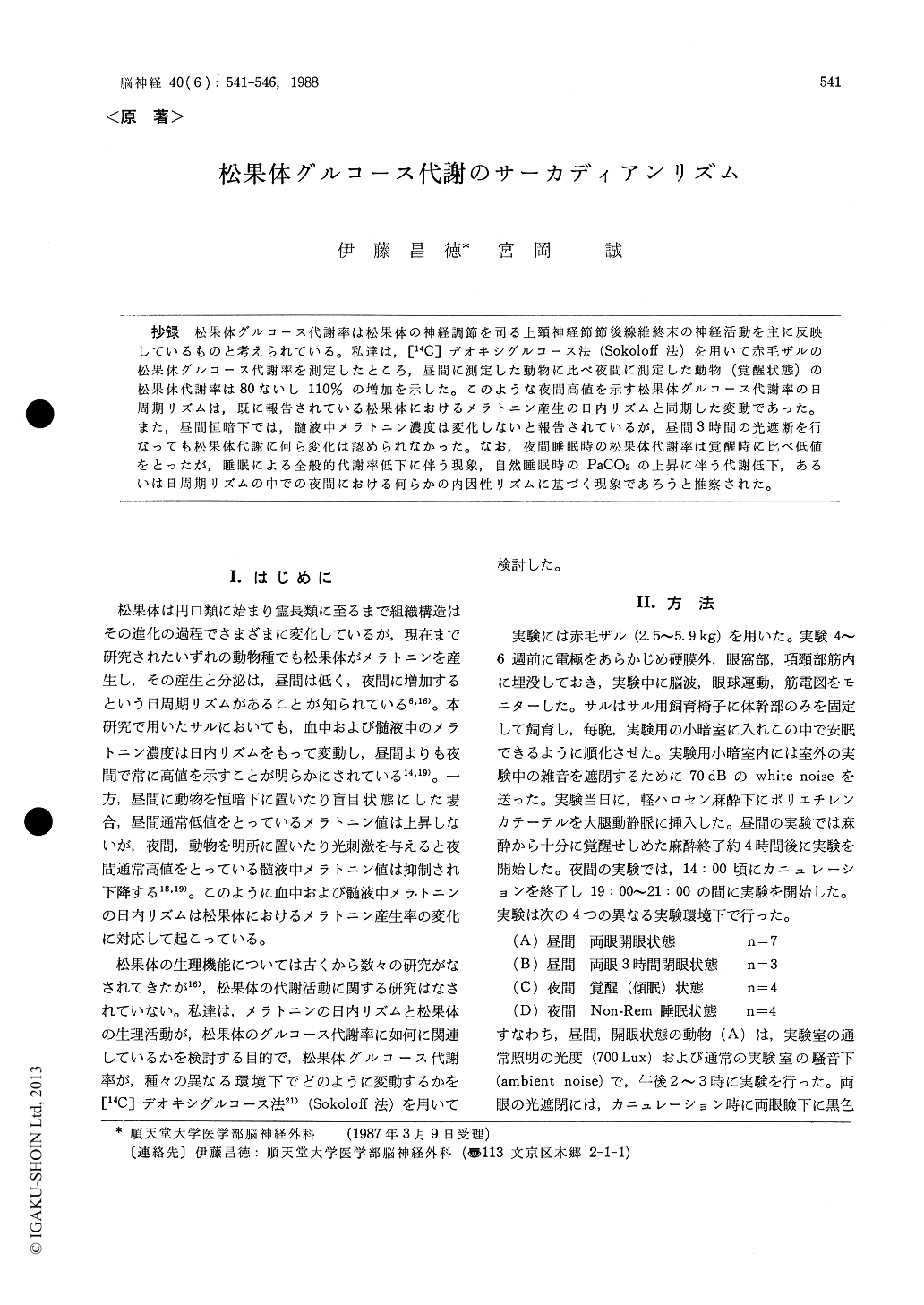
抄録 松果体グルコース代謝率は松果体の神経調節を司る上頸神経節節後線維終末の神経活動を主に反映しているものと考えられている。私達は,[14C]デオキシグルコース法(Sokoloff法)を用いて赤毛ザルの松果体グルコース代謝率を測定したところ,昼間に測定した動物に比べ夜間に測定した動物(覚醒状態)の松果体代謝率は80ないし110%の増加を示した。このような夜間高値を示す松果体グルコース代謝率の日周期リズムは,既に報告されている松果体におけるメラトニン産生の日内リズムと同期した変動であった。また,昼間恒暗下では,髄液中メラトニン濃度は変化しないと報告されているが,昼間3時間の光遮断を行なっても松果体代謝に何ら変化は認められなかった。なお,夜間睡眠時の松果体代謝率は覚醒時に比べ低値をとったが,睡眠による全般的代謝率低下に伴う現象,自然睡眠時のPaCO2の上昇に伴う代謝低下,あるいは日周期リズムの中での夜間における何らかの内因性リズムに基づく現象であろうと推察された。
We employed the quantitative 2-〔14C〕-deoxyglu-cose method (Sokoloff's method) to measure glucose utilization in the pineal gland of pubescent mon-keys. Glucose utilization in the pineal was 80-110 % higher in the nocturnal, awake animals com-pared to the rate of both groups studied in the noc-turnal, awake animals with both eyes open and with light deprivation for three hours. Short term visual deprivation during the day was without effect on pineal glucose utilization.
The diurnal variations in melatonin levels in blood and CSF, higher at night than during the day, are the result of corresponding changes in the rate of production and elaboration of melatonin in the pineal galnd. The release of norepinephrine from the postganglionic fiber of the superior cer-vical ganglia controls the production of melatonin in the pineal by regulating the activity of seroto-nin-N-acetyltransferase. It was reported that elect-rical stimulation of SCG via sympathetic trunk increased the levels of serotonin-N-acetyltrans-ferase in the pineal and that it also increased glucose utilization in the pineal. It is believed that meta-bolic increase in the pineal reflects increased acti-vity in sympahtetic terminals distributed through-out the gland which stimulate its increase in hor-mone production. The present results indicate that there is an elevation of pineal metabolic rate at a time when blood and CSF levels of melatoninare known to be elevated. Our finding that short-term light deprivation during the day did not af-fect the pineal metabolic rate is consistent with the result by Reppert et al (1981) in which they found that exposure to darkness during the day does not result in an increase in CSF melatonin.
This work was supported by National Institutes of Health (USA) visiting program. M. I. and M. M. were the visiting associates at Laboratory of Cerebral Metabolism, N. I. M. H.. The authors appreciate advice and support of Drs. Louis Sokoloff and Charles Kennedy, LCM, NIMH, and Dr. Richard Nakamura, Laboratory of Psychology and Psychopathology, NIMH.

Copyright © 1988, Igaku-Shoin Ltd. All rights reserved.


