Japanese
English
- 有料閲覧
- Abstract 文献概要
- 1ページ目 Look Inside
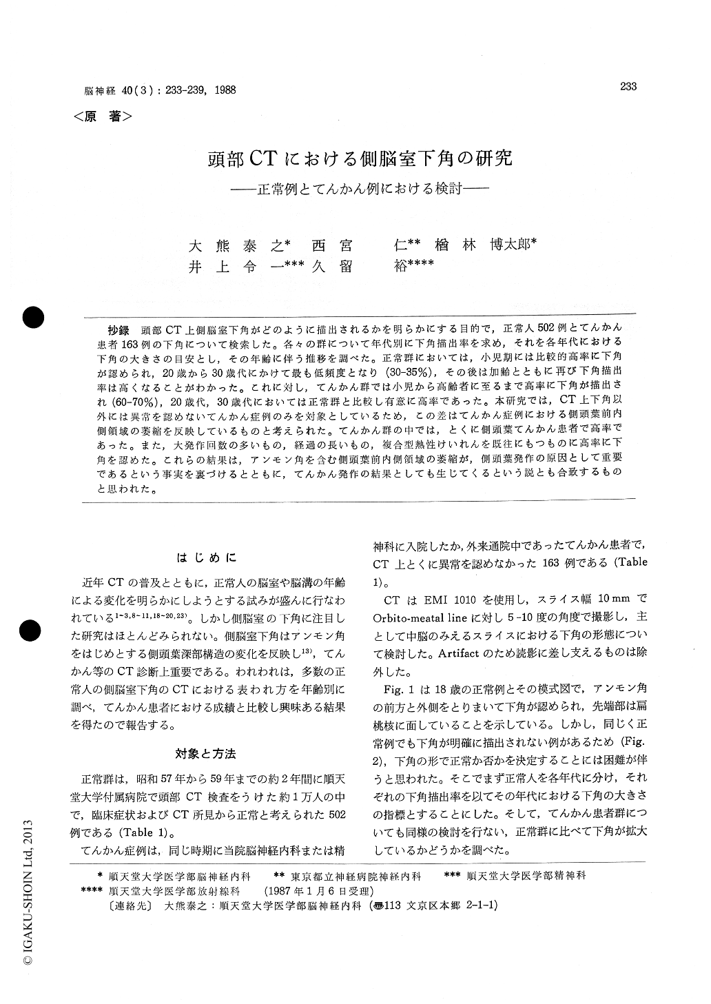
抄録 頭部CT上側脳室下角がどのように描出されるかを明らかにする目的で,正常人502例とてんかん患者163例の下角について検索した。各々の群について年代別に下角描出率を求め,それを各年代における下角の大きさの目安とし,その年齢に伴う推移を調べた。正常群においては,小児期には比較的高率に下角が認められ,20歳から30歳代にかけて最も低頻度となり(30-35%),その後は加齢とともに再び下角描出率は高くなることがわかった。これに対し,てんかん群では小児から高齢者に至るまで高率に下角が描出され(60-70%),20歳代,30歳代においては正常群と比較し有意に高率であった。本研究では,CT上下角以外には異常を認めないてんかん症例のみを対象としているため,この差はてんかん症例における側頭葉前内側領域の萎縮を反映しているものと考えられた。てんかん群の中では,とくに側頭葉てんかん患者で高率であった。また,大発作回数の多いもの,経過の長いもの,複合型熱性けいれんを既往にもつものに高率に下角を認めた。これらの結果は,アンモン角を含む側頭葉前内側領域の萎縮が,側頭葉発作の原因として重要であるという事実を裏づけるとともに,てんかん発作の結果としても生じてくるという説とも合致するものと思われた。
In recent years brain CT scan has been so popular that many investigators have been trying to clarify the normal CT images. But little atten-tion has been paid to the inferior horns of lateral ventricles in spite of their importance for judg-ing mesial temporal lobe structures. The present study was designed to elucidate how the inferior horns were visualized on brain CT in normal subjects from childhood to aged group, and to evaluate whether the inferior horns were dilated in the epileptics or not.
The subjects of the present study were 502 normal controls (2-79 y, mean 36.1 y) and 163 epileptic patients with normal CT image (4-68 y, mean 26.6 y) including 55 cases of temporal lobe epileptics. CT scans were performed with EMI 1010 scanner, and slices were obtained every 10 mm from the projection of 5-10° angle for orbito-meatal line. Inferior horns were examined at the level of basal cisterns. Because inferior horns were not necessarily visible in our cases, we examined the frequency of clearly visualized inferior horns at each decade, and regarded their frequency as the size of inferior horns at each decade.
In normal controls, frequency of visible inferior horns was relatively high in early childhood, and decreased as they grew. In the 3 rd to 4 th decades the visibility frequency became the lowest (30-35%), and then gradually increased as the subjects became older. The reason of high fre-quency in children was not explained, but in-creased frequency in the aged might be due to generalized atrophy because of their parallelism to increasing ventricular indices (i. e. bicaudate index and inverse cella media index).
In epileptic patients, in contrast, inferior horns were highly visible (60-70%) in all generations from childern to aged patients. The difference of frequency between epileptics and normal controls was statistically significant in 3 rd and 4 th de-cades, and indicates that inferior horns in epilep-tics are more larger than in the normal controls.
And this is explained as due to atrophy of the mesial temporal structures including Ammon's horn in epileptics.
According to the classification of epilepsy, temporal lobe epileptics had especially highly visible inferior horns. This may be in agreement with the consensus that Ammon's horn sclerosis is one of the major cause of temporal lobe sei-zure. On the other hand, inferior horns were highly visualized in epileptics with frequent grand mal seizures, with histories of comlpex type febrile convulsions and with long-standing course. These findings also may suggest that atrophy of mesial temporal structures is partially due to anoxia resulting from repeated seizures.

Copyright © 1988, Igaku-Shoin Ltd. All rights reserved.


