Japanese
English
- 有料閲覧
- Abstract 文献概要
- 1ページ目 Look Inside
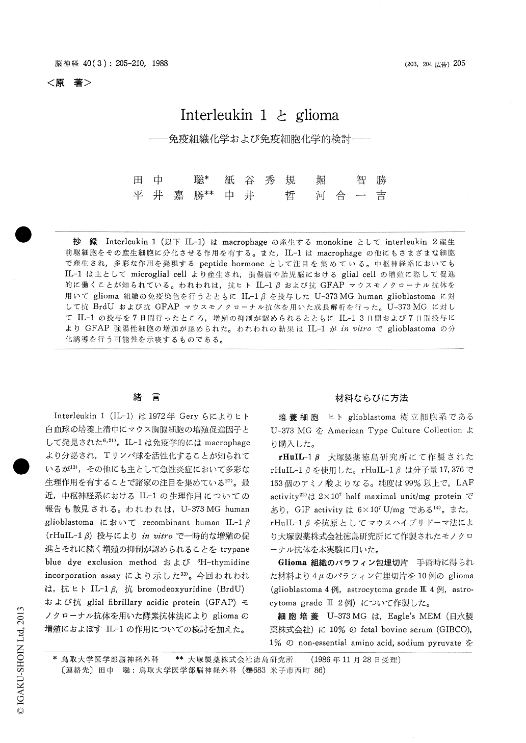
抄録 Interleukin1(以下IL−1)はmacrophageの産生するmonokineとしてinterleukin2産生前駆細胞をその産生細胞に分化させる作用を有する。また,IL−1はmacrophageの他にもさまざまな細胞で産生され,多彩な作用を発現するpeptide hormoneとして注目を集めている。中枢神経系においてもIL−1は主としてmicroglial cellより産生され,損傷脳や胎児脳におけるglial cellの増殖に際して促進的に働くことが知られている。われわれは,抗ヒトIL−1βおよび抗GFAPマウスモノクローナル抗体を用いてglioma組織の免疫染色を行うとともにIL−1βを投与したU−373MG human glioblastomaに対して抗BrdUおよび抗GFAPマウスモノクローナル抗体を用いた成長解析を行った。U−373MGに対してIL−1の投与を7日間行ったところ,増殖の抑制が認められるとともにIL−13日間および7日間投与によりGFAP強陽性細胞の増加が認められた。われわれの結果はIL−1がin vitroでglioblastomaの分化誘導を行う可能性を示唆するものである。
Interleukin 1 is a monokine produced by mac-rophage and has an ability to activate thymo-cytes. In addition to the immunological regulatory effect, interleukin 1 has attracted a great deal of invetsigators as a new peptide hormon that was secreted by many cells and has a various physio-logical activities. In central nervous systems, in-terleukin 1 promotes the glial cells proliferations on the injured brain and the fetal brain. The cell sourses of interleukin 1 in central nervous systems are considered to the microglial cells.
On gliomas, Lachmann and Dinarello reported growth promoting effect of IL-1 on U-373 MG human glioblastoma cell. The authors investigated the roles and effects of IL-1 on the growth of gliomas using recombinant human IL-1 β and anti-HuIL-1 β monoclonal antibody.
On Immunohistochemistry, paraffin sections of 10 cases of gliomas were stained with immunoper-oxidase method using anti-human IL-1 β and anti-GFAP mouse monoclonal antibody. All astro-cytomas examined and 2 of 4 glioblastomas were stained by anti-IL-1β. The origin of IL-1 that was stained by immunoperoxidase staining is un-known. The authors think it that IL-1 existed in glioma cells were secreted by microglial cells or that the glioma cells themselves secreted IL-1. In either case, IL-1 must be related to the growth of glioma in situ.
On immunocytochemistry, U-373 MG human glioblastoma cells purchased from ATCC were incubated on cover-slip with 0 and 10 U/ml of rHuIL-1 β for 3 or 7 days. The cells were stained with immunoperoxidase method using anti-GFAP monoclonal antibody. The marked increase of GFAP-positive cells were observed in IL-1 treated U-373 MG glioblastoma cells.
The authors already reported that rHuIL-1 β given in vitro to U-373 MG cell had caused a transient promotion of cellular growth followed by inhibition. Nagashima and Hoshino successfully employed anti-BrdU monoclonal antibody to study cell kinetics of brain tumors. According to these procedures, U-373 MG incubated with 10 U/ml of rHuIL-1 β for 0, 3 and 7 days were treated with 5 μM BrdU for 30 minutes. The cultured cells were stained on cover-slip with immunoperoxidase method using anti-BrdU mouse monoclonal anti-body. The marked decrease of BrdU-positive cells was observed in IL-1 treated U-373 MG for 7 days.
Presumably, IL-1 has a possibility to induce the differentiation of glioblastoma, considering its causing the transient growth facilitation followed by inhibition when given in vitro to human glioblastoma cells. The increase of GAFP-stain-ability of U-373 MG treated with rHuIL-1 β par-tially confirmed this hypothesis.

Copyright © 1988, Igaku-Shoin Ltd. All rights reserved.


