Japanese
English
- 有料閲覧
- Abstract 文献概要
- 1ページ目 Look Inside
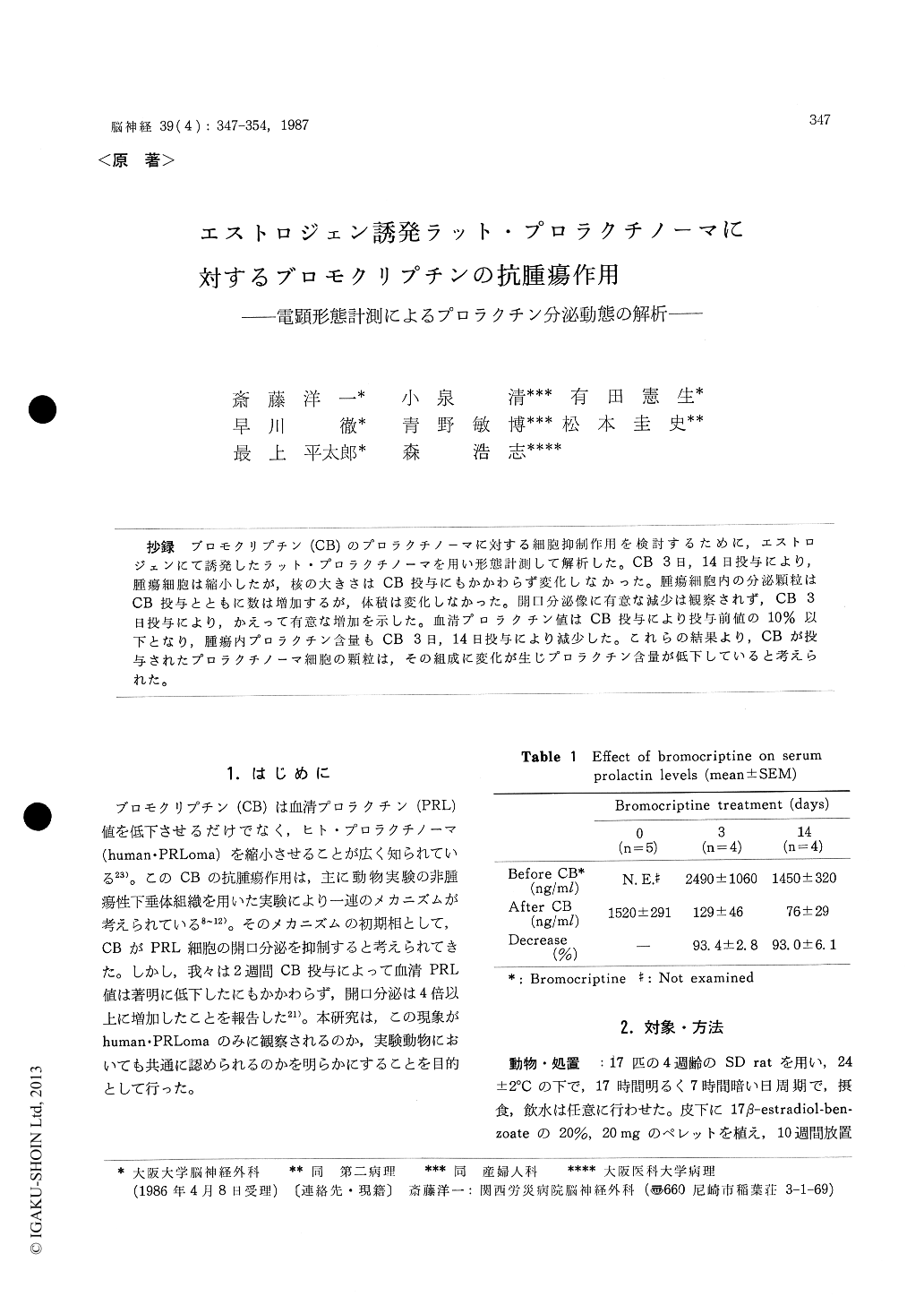
抄録 プロモクリプチン(CB)のプロラクチノーマに対する細胞抑制作用を検討するために,エストロジェンにて誘発したラット・プロラクチノーマを用い形態計測して解析した。CB 3日,14目投与により,腫瘍細胞は縮小したが,核の大きさはCB投与にもかかわらず変化しなかった。腫瘍細胞内の分泌顆粒はCB投与とともに数は増加するが,体積は変化しなかった。開口分泌像に有意な減少は観察されず,CB 3日投与により,かえって有意な増加を示した。血清プロラクチン値はCB投与により投与前値の10%以下となり,腫瘍内プロラクチン含量もCB 3日,14日投与により減少した。これらの結果より,CBが投与されたプロラクチノーマ細胞の顆粒は,その組成に変化が生じプロラクチン含量が低下していると考えられた。
Bromocriptine (CB) not only lowers serum prolactin (PRL) levels but also reduces tumor size of human prolactinomas. Gen et al and we have suggested that the size reduction of human prolactinomas by bromocriptine treatment results from the reduction in size of individual tumor cell as well as the reduction in number of tumor cells secondary to cell necrosis. This implies that bromocriptine has a cytosuppressive action and possibly a cytocidal action on human prolactinomaswhich causes reduction in cell size and cell ne-crosis, respectively. The mechanism of cytosuppres-sive action of CB has been investigated by using mostly non-neoplastic pituitary tissues of experi-mental animals. A decrease in exocytosis of secre-tory granules and a subsequent accumulation of granules within the cells are suggested to cause the reduction in serum levels of PRL in early stage of CB treatment. However we have reported that in spite of a pronounced reduction of serum PRL levels, the number of exocytosis of the gra-nules in human prolactinomas treated with CB for 2 weeks increased to more than 4 times much as that in the untreated prolactinomas. This is a phenomenon which contradictory to the current hypothesis. The present study is intended to cla-rify whether the phenomenon we observed is specific for human prolactinomas or common also to the prolactinomas in experimental animals.
Seventeen female SD rats were used. They were implanted subcutaneously with a pellet of 20 mg of 17 estradiol-benzoate (20% in cholesterol), and left to grow a pituitary tumor for 10 weeks. Immunohistochemistry at light microscopic level revealed that more than 90% of the tumor cells were positive for rat PRL. There was no definite necrosis or destruction of tumor cells in any tumor examined. The tumor cells treated with CB for 3 or 14 days were smaller in size than those in control group. However, the nuclei of tumor cells in CB treated and no treated tumors were almost the same in size. A quantitative analysis of ul-trastructural alterations of secretory granules in correlation with PRL concentrations in estrogen-induced rat prolactinomas was performed to inves-tigate mechanisms of a cytosuppressive action of CB. The secretory granules of CB-treated adeno-mas increased in number but not in volume. The number of exocytosis of the granules showed no significant decrease, but rather an increase in adenomas treated with CB for 3 days. Radio-immunoassay revealed a decrease in PRL concen-trations of adenoma tissues, and a marked reduc-tion in serum PRL levels. These results suggest a little amount of PRL in secretory granules of the CB-treated pituitary adenomas.

Copyright © 1987, Igaku-Shoin Ltd. All rights reserved.


