Japanese
English
- 有料閲覧
- Abstract 文献概要
- 1ページ目 Look Inside
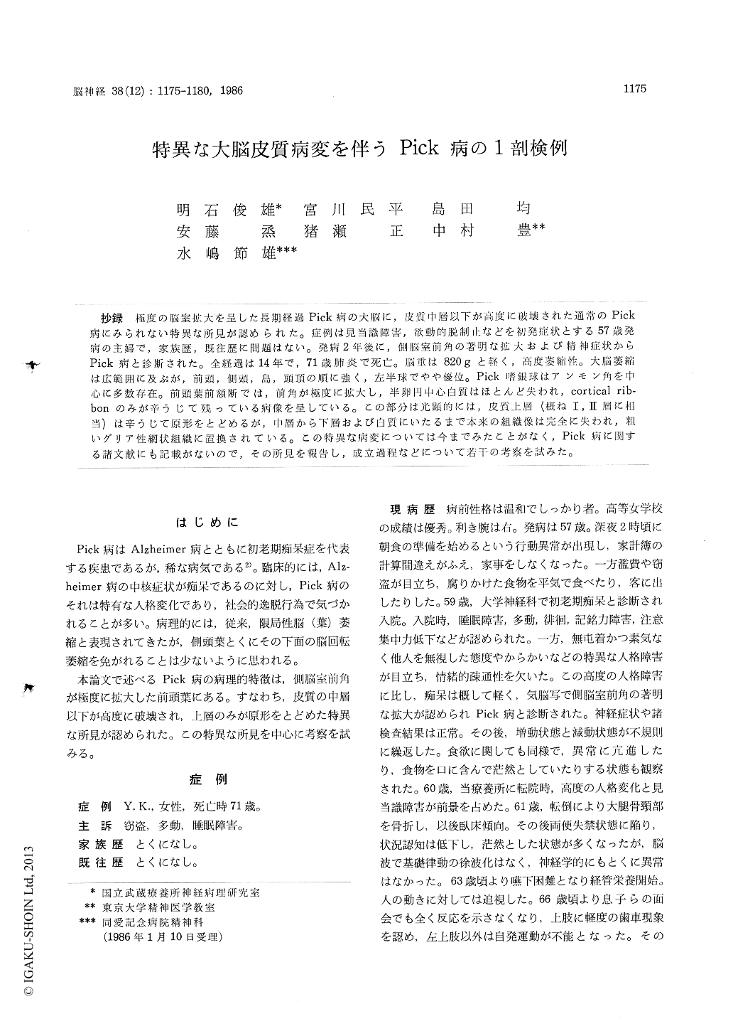
抄録 極度の脳室拡大を呈した長期経過Pick痛の大脳に,皮質中層以下が高度に破壊された通常のPick病にみられない特異な所見が認められた。症例は見当識障害,欲動的脱制止などを初発症状とする57歳発病の主婦で,家族歴,既往歴に問題はない。発病2年後に,側脳室前角の著明な拡大および精神症状からPick病と診断された。全経過は14年で,71歳肺炎で死亡。脳重は820gと軽く,高度萎縮性。大脳萎縮は広範囲に及ぶが,前頭,側頭,島,頭頂の順に強く,左半球でやや優位。Pick嗜銀球はアンモン角を中心に多数存在。前頭葉前額断では,前角が極度に拡大し,半卵円中心白質はほとんど失われ,cortical rib-bonのみが辛うじて残っている病像を呈している。この部分は光顕的には,皮質上層(概ねI,II層に相当)は辛うじて原形をとどめるが,中層から下層および白質にいたるまで本来の組織像は完全に失われ,粗いグリア性網状組織に置換されている。この特異な病変については今までみたことがなく,Pick病に関する諸文献にも記載がないので,その所見を報告し,成立過程などについて若干の考察を試みた。
An autopsied case of Pick's disease, having an extraordinary cerebral change in the anterior port-tion of Lobus frontalis and temporalis, was report ed.
Our case is a 71 year-old woman at death with a fourteen year history of chronic progressive dementia and mental deterioration, and it may be stressed that the existence lasted 8 years, over thg latter half of clinical course, was depended on the tube feeding. The first symptoms suddenly appeared in 1964, 2 months after her husband's death of illness, when she was 57. She prepared the table for breakfast late at night, calculated wrongly in her domestic account book, and stole foods in the grocery.
Two years later, her illness was diagnosed as presenile dementia by characteristic personality change and marked dilatation of anterior horn of lateral ventriculus.
On admission to National Musashi Sanatorium, three years after the first symptoms' appearrance, she presented restless walking, insomnia, memory loss, weakness of concentration, and high degree of disorientation. Particularly, it was noticeable that she behaved with bizzare contact.
After 1970, tube feeding was introduced conti-nuously, because of swallowing difficulty. Death occured in July 1978 from a general weakness and a broncho-pneumonia, 14 years after the onset of the first symptoms.
Autopsy revealed small and atrophied brainweighed 820 g. Cerebral cortical atrophy extended to frontal, temporal, insular, and parietal lobes, but right T-1 was relatively well preserved. On sec-tion, frontal and temporal ventriculus were remar-kably enlarged and caudate nuclei were extremely atrophic. Although, Alzheimer's neurofibrillary changes and senile plaques were scarcely recog-nized, but Pick's bodies (argyrophilic inclusions) were widely distributed to the atrophied cerebral cortices, particularly numerous in the Ammon's horn.
Ultrastructurally, a Pick's body was composed of osmiophilic aggregates, mitochondria, vesicles, and a few filaments which seemed microtubules of 130-150 Å in diameter.
By the way, neuropathological characteristic of this case lie in the cortex of frontal and temporal lobes, as we discribed at the beginning. In that regions, macroscopically, cortical ribbon was very thin (Fig. 2), and more deep portion including white matter was hardly been stained by any me-thods of HE, KB, Heidenhain-Woelcke, Holzer and so on. Microscopically, this unstainable region did correspond to deeper layers (III~VI) and white matter (Fig. 3 A). In that region, cytoarchitecto-nics was completely destroyed and replaced with rough glial fiber networks in which Pick's bodies were scattered (Fig. 4), and original border of cortex and white matter was disappered.
We considered that this cerebral change did not belong to Pick's atrophic process, but, the regions mentioned above coincided with that of early and yet predilective portion for Pick's cerebral atrophy, so it was impossible for us to come a conclusion.
This cerebral change could hardly be find in Pick's reports, but resemblant findings were barely found in reports of hyperammonemia, although the clinical symptoms involving laboratory data dif-fered quite from that of Pick's disease.
We assume that this cerebral change was ad-vanced within the period of time that the patient survived in so-called vegetative state depending on tube feeding, but true cause is still unknown.

Copyright © 1986, Igaku-Shoin Ltd. All rights reserved.


