Japanese
English
- 有料閲覧
- Abstract 文献概要
- 1ページ目 Look Inside
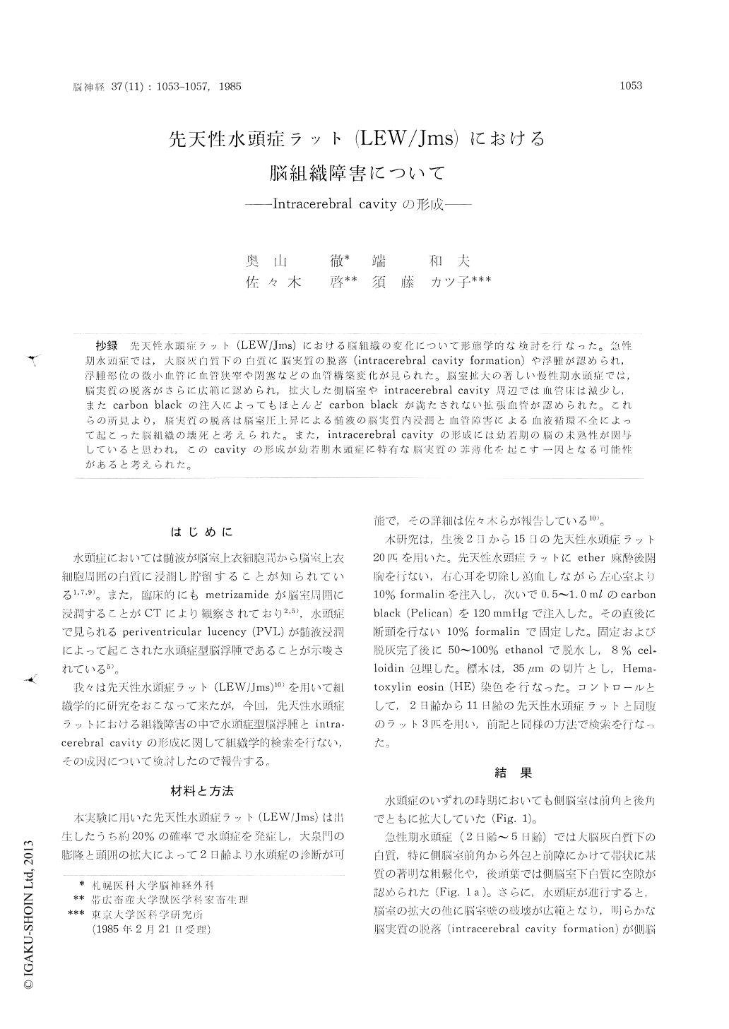
抄録 先天性水頭症ラット(LEW/Jms)における脳組織の変化について形態学的な検討を行なった。急性期水頭症では,大脳灰白質下の白質に脳実質の脱落(intracerebral cavity formation)や浮腫が認められ,浮腫部位の微小血管に血管狭窄や閉塞などの血管構築変化が見られた。脳室拡大の著しい慢性期水頭症では,脳実質の脱落がさらに広範に認められ,拡大した側脳室やintracerebral cavity周辺では血管床は減少し,またcarbon blackの注入によってもほとんどcarbon blackが満たされない拡張血管が認められた。これらの所見より,脳実質の脱落は脳室圧上昇による髄液の脳実質内浸潤と血管障害による血液循環不全によって起こった脳組織の壊死と考えられた。また,intracerebral cavityの形成には幼若期の脳の未熟性が関与していると思われ,このcavityの形成が幼若期水頭症に特有な脳実質の菲薄化を起こす一因となる可能性があると考えられた。
Brain tissue damage in congenital hydrocephalic rat (LEW/Jms) was studied in the aspects of the hydrocephalic brain edema and the changes of the vascular apparatus. The characteristic findings in this study were the changes of the small blood vessels and the intracerebral cavity formation.
In the acute stage of hydrocephalus (2 to 5 days after birth), spongy appearance and necrosisof the brain edema were observed in the periven-tricular white matter. The stenotic or obstructive vascular changes were located in connection with the hydrocephalic brain tissue. In this stage, the intracerebral cavity was formed particularly in the periventricular edematous white matter result-ing in a thinning of the occipital lobes.
In the late stage of hydrocephalus (9 to 15 days after birth), the lateral ventricles were severely dilated, and a markedly dilatated intracerebral cavity was observed in the periventricular white matter. The edematous area was observed adjacent to the dilated lateral ventricles or the intracere-bral cavity. In the late stage, the number of small vessels filled with carbon black decreased in the area of the CSF edema when compared to the acute stage, and many obstructive blood vessels were observed in the same area. Moreover, dila-tated blood vessels without carbon black were observed in the border zone between the normal and the edematous area adjacent to the intra-cerebral cavity.
These vascular changes may occur by the ac-cumulation of the CSF as well as the mechanical compression, and consequently lead to the micro-circulatory disturbance. These microcirculatory disturbances may contribute to the intracerebral cavity formation with the accumulation of the CSF in the extracellular space.
The thinning of the brain tissue observed in congenital hydrocephalus may be the result of the intracerebral cavity formation that could occur only in the immature brain tissue.

Copyright © 1985, Igaku-Shoin Ltd. All rights reserved.


