Japanese
English
- 有料閲覧
- Abstract 文献概要
- 1ページ目 Look Inside
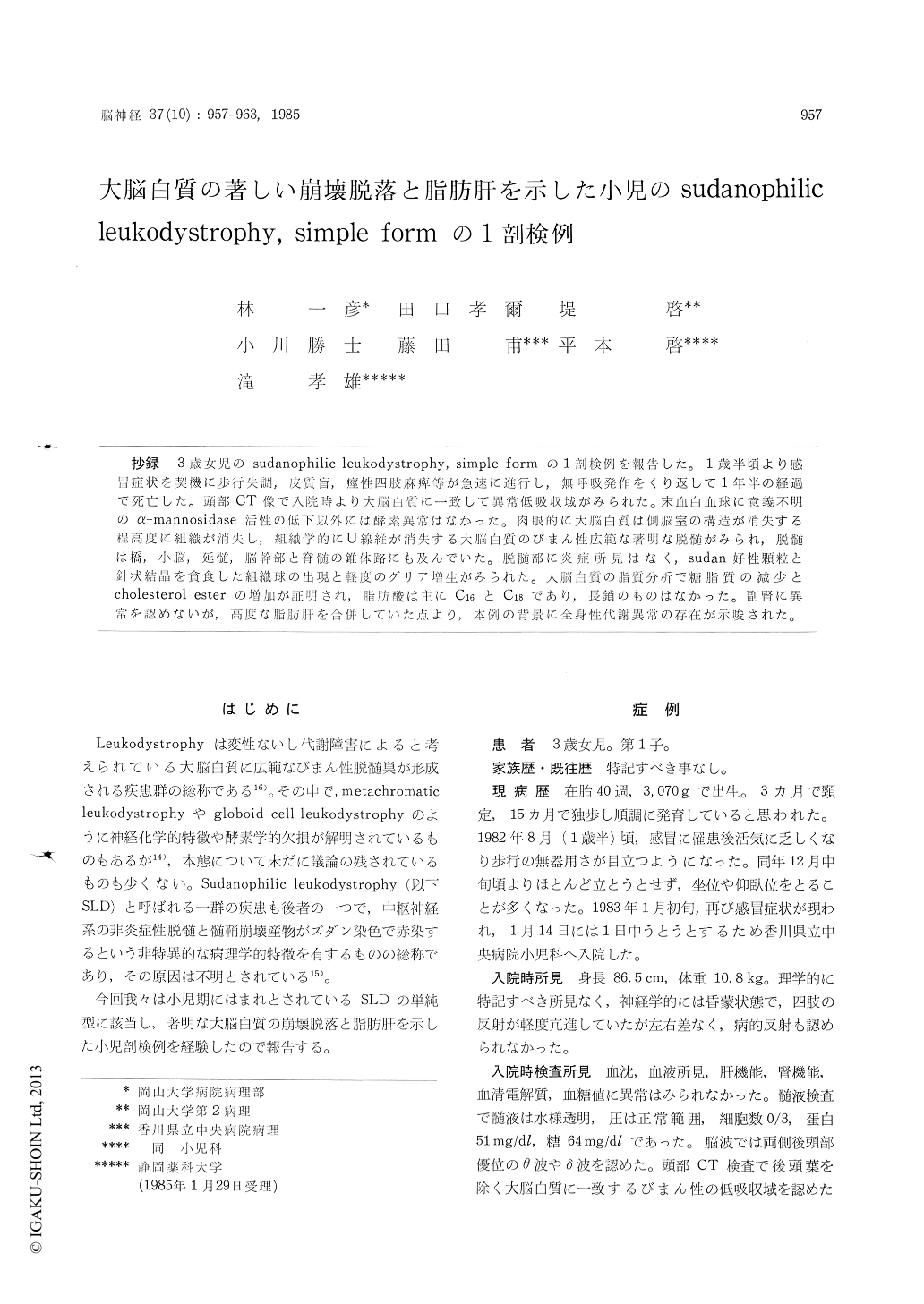
抄録 3歳女児のsudanophilic leukodystrophy, simple formの1剖検例を報告した。1歳半頃より感冒症状を契機に歩行失調,皮質盲,痙性四肢麻痺等が急速に進行し,無呼吸発作をくり返して1年半の経過で死亡した。頭部CT像で入院時より大脳白質に一致して異常低吸収域がみられた。末血白血球に意義不明のα—mannosidase活性の低下以外には酵素異常はなかった。肉眼的に大脳白質は側脳室の構造が消失する程高度に組織が消失し,組織学的にU線維が消失する大脳白質のびまん性広範な著明な脱髄がみられ,脱髄は橋,小脳,延髄,脳幹部と脊髄の錐体路にも及んでいた。脱髄部に炎症所見はなく,sudan好性顆粒と針状結晶を貪食した組織球の出現と軽度のグリア増生がみられた。大脳白質の脂質分析で糖脂質の滅少とcholesterol esterの増加が証明され,脂肪酸は主に C16 と C18 であり,長鎖のものはなかった。副腎に異常を認めないが,高度な脂肪肝を合併していた点より,本例の背景に全身性代謝異常の存在が示唆された。
A sporadic case of sudanophilic leukodystrophy of the simple form (Peiffer) was reported. The patient was three-year-old girl who had suffered from progressive developmental retardation and neurological disorders such as ataxia, cortical blind-ness and spastic paralysis of the extremities for eighteen months after she had showed normal development till one and a half years old and died from respiratory insufficiency. On admission, computerized tomogram scan demonstrated diffuse low density lesions of the cerebral white matter extending subsequently to the subcortical white matter. Examination of cerebrospinal fluid reveal-ed only slight increase of protein. Lysosomal enzyme activities such as arylsulfatase and β- galactosidase in the white blood cells were normal except for distinctly low activity of a-mannosidase without any clinical symptoms suggesting a-man-nosidase deficiency. Amino acids in blood were normal.
The brain weighed 900 gm. On the coronal sec-tions most part of the cerebral white matter was so strongly degenerated and disappeared that the lateral ventricular structure was not discernible. Histologically, a diffuse and symmetrical demylina-tion, loss of axons including U fibers and mode-rate gliosis were observed in the residual white matter in the cerebrum and pons. There was no inflammatory cells and metachromatic substances. Large amount of sudanophilic droplets showing polarizing cross and needle like crystals were found in the intra- and/or extracytoplasm of macropha-ges. Demyelinated lesions with little tissue reac-tion were also found in the cerebellum, medulla oblongata and in pyramidal tracts through mid-brain to cervical spinal cord. There were slight loss of neurons and moderate astrocytosis in the cerebral cortex and basal ganglia. There were no Rosenthal fibers and no sparing of islets of myelin.
Lipid analysis of the formalin-fixed cerebral white matter revealed decrease of cholesterol, sulfatide and galactosylceramide and increase of esterified cholesterol. Fatty acids of C: 16 and C: 18 were overwhelmingly dominant components but no long chain fatty acids were detected in cho-lesterol ester.
There was no lesion in the adrenal glands and no membranocystic degeneration but the liver showed a marked diffuse fatty degeneration despite no diabetes mellitus and malnutrition which sug-gests the possibility that this case might have a systemic disorder of abnormal lipid metabolism as the background, like as adrenoleukodystrophy.

Copyright © 1985, Igaku-Shoin Ltd. All rights reserved.


