Japanese
English
- 有料閲覧
- Abstract 文献概要
- 1ページ目 Look Inside
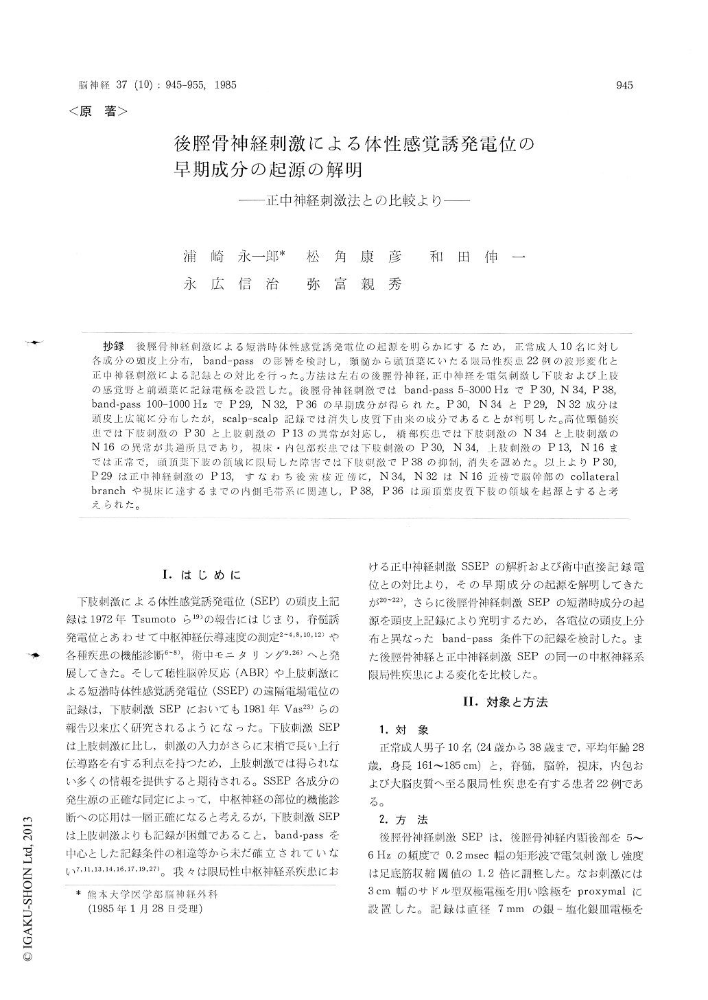
抄録 後脛骨神経刺激による短潜時体性感覚誘発電位の起源を明らかにするため,正常成人10名に対し各成分の頭皮上分布,band-passの影響を検討し,頸髄から頭頂葉にいたる限局性疾患22例の波形変化と正中神経刺激による記録との対比を行った。方法は左右の後脛骨神経,正中神経を電気刺激し下肢および上肢の感覚野と前頭葉に記録電極を設置した。後脛骨神経刺激ではband-pass 5-3000 HzでP 30,N 34,P 38,band-pass 100-1000 HzでP29,N 32,P 36の早期成分が得られた。P 30,N 34とP29,N 32成分は頭皮上広範に分布したが,scalp-scalp記録では消失し皮質下由来の成分であることが判明した。高位頸髄疾患では下肢刺激のP 30とL肢刺激のP 13の異常が対応し,橋部疾患では下肢刺激のN 34と上肢刺激のN 16の異常が共通所見であり,視床・内包部疾患では下肢刺激のP 30,N 34,上肢刺激のP 13,N 16までは正常で,頭頂葉下肢の領域に限局した障害では下肢刺激でP 38の抑制,消失を認めた。以上よりP 30,P 29は正中神経刺激のP 13,すなわち後索核近傍に,N 34,N 32はN 16近傍で脳幹部のcollateralbranchや視床に達するまでの内側毛帯系に関連し,P 38,P 36は頭頂葉皮質下肢の領域を起源とすると考えられた。
In order to determine the generation sites of short latency somatosensory evoked potentials to the posterior tibial nerve stimulation, scalp topo-graphy was performed on 10 normal subjects in the two different band-pass recordings, i. e., wide band-pass filter (5-3000 Hz) and narrow band-pass filter (100-1000 Hz). Furthermore, comparative study of the changes of evoked potentials between posterior tibial nerve stimulation and median ner-ve stimulation was carried out in 22 cases with well localized lesion of the central nervous system in the same wide band-pass filter setting.
The early components of somatosensory evoked potentials elicited by the posterior tibial nerve stimulation were obtained as P 30, N 34, and P 38 in the wide band-pass filter, and P 29, N 32, P 36 in the narrow band-pass filter. Components P 30, N 34 and components P 29, N 32 were widely dis-tributed on the scalp, but were disappeared on the scalp-scalp recording. These results suggested all those components were generated from the deep subcortical structures.
In the case with high cervical lesion, component P 30 at the posterior tibial nerve stimulation was remarkably prolonged in latency, and component P 13 at the median nerve stimulation was disap-peared. P 30-N 34 interpeak latency at the pos-terior tibial nerve stimulation was prolonged in the case with pontine lesion, while P 13-N 16 inter-peak latency at the median nerve stimulation was also prolonged. In the cases with thalamic and in-ternal capsular lesion, P 30 and N 34 at the pos-terior tibial nerve stimulation and P 13 and N 16 at the median nerve stimulation were all preser-ved in normal range. These results revealed that components P 30 and N 34 were almost identical to components P 13 and N 16, respectively. On the other hand, component P 38 at the posterior tibial nerve stimulation was suppressed or disappeared in the cases with well localized lesion at the mid-centro parietal region, that includes the primary foot sensory area.
In conclusion, the generation sites of each early component of somatosensory evoked potentials to posterior tibial nerve stimulation were considered as follows; a) components P 30 and P 29 were almost same to the component P 13 at the median nerve stimulation that was generated from near by the dorsal column nucleus, b) components N 34 and N 32 were similar to the component N 16 atthe median nerve stimulation that reflected the activities of the collateral branch from the medial lemniscus at the brain stem and/or the afferent volley of the medial lemniscal pathway between the dorsal column nucleus and the thalamus, c) components P 38 and P 36 were originated from the primary foot sensory area at the parietal lobe.

Copyright © 1985, Igaku-Shoin Ltd. All rights reserved.


