Japanese
English
- 有料閲覧
- Abstract 文献概要
- 1ページ目 Look Inside
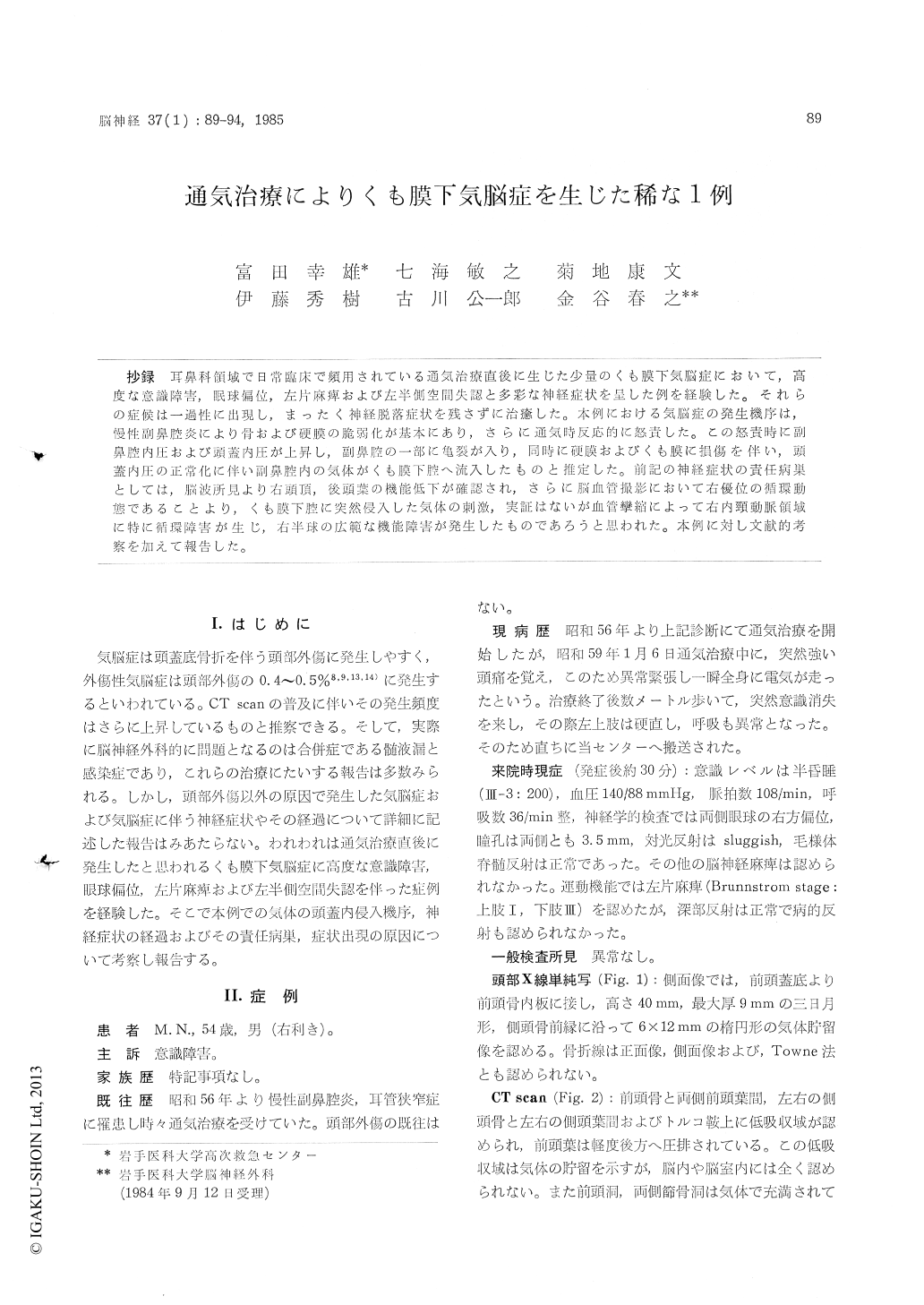
抄録 耳鼻科領域で日常臨床で頻用されている通気治療直後に生じた少量のくも膜下気脳症において,高度な意識障害,眠球偏位,左片麻痺および左半側空間失認と多彩な神経症状を呈した例を経験した。それらの症候は一過性に出現し,まったく神経脱落症状を残さずに治癒した。本例における気脳症の発生機序は,慢性副鼻腔炎により骨および硬膜の脆弱化が基本にあり,さらに通気時反応的に怒責した。この怒責時に副鼻腔内圧および頭蓋内圧が上昇し,副鼻腔の一部に亀裂が入り,同時に硬膜およびくも膜に損傷を伴い,頭蓋内圧の正常化に伴い副鼻腔内の気体がくも膜下腔へ流入したものと推定した。前記の神経症状の責任病巣としては,脳波所見より右頭頂,後頭葉の機能低下が確認され,さらに脳血管撮影において右優位の循環動態であることより,くも膜下腔に突然侵入した気体の刺激,実証はないが血管攣縮によって右内頸動脈領域に特に循環障害が生じ,右半球の広範な機能障害が発生したものであろうと思われた。本例に対し文献的考察を加えて報告した。
No detailed reports on pneumocephalus caused by any factors other than head trauma, and their courses accompanied with this disease have been so far available. We recently experienced a case of pneumocephalus complicated with severe cloud-ing of consciousness, ocular deviation, and unilateral spatial neglect with the results being reported hereinafter.
A 54-year-old man had often received ear douche therapy due to chronic sinusitis and tubal obstrution about 3 years before without any history of head trauma. On Jan. 6, 1984, sudden clouding of consciousness accompanying stiffness of the left arm occurred immediately after ear douche, and then he was transferred to our center. At admission, semicomatose, bilateral ocular devia-tion to the right, and left hemiparesis were observed. Plain skull X-ray films showed a reten-tion of air in the frontal and temporal regions, while CT scan revealed air retention on the bilateral frontal region, bilateral temporal tip and suprasellar cistern. However, no abnormal findings were detected in the brain. Consiciousness and hemiparesis recovered on the next day of hospita-lization, however, the left hemispatial neglect still remained. This symptom was still observed on the 3rd day but disappeared by the 4th day of hospitalization.
For clarifying its cause, cerebral angiography, CT scan and electroencephalography were then performed. CT scan revealed no anomalies in the brain, while cerebral angiography showed a cere-bral circulation patttern in favor of the right internal carotid artery. In the electroencephalo-gram, the background activity was consisted of a-wave, but the right pattern was irregular, especially complicated with slow a and 0 waves in the parietal and occipital leads. These findings suggested a decreased cerebral function and theleft hemispatial neglect were considered to be of symptoms caused by the right parietal and occipital lesions, i, e., the circulation pattern in favor of the right sided arteries. Thus, probably due to air stimulation invaded into the subarachroideal cavity and to subsequent vasospasm, disturbed circulation in the right carotid artery might pro-voke the appearance of neurologic symptoms and functional disturbance in the right hemisphere noted in the electroencephalogram.

Copyright © 1985, Igaku-Shoin Ltd. All rights reserved.


