Japanese
English
- 有料閲覧
- Abstract 文献概要
- 1ページ目 Look Inside
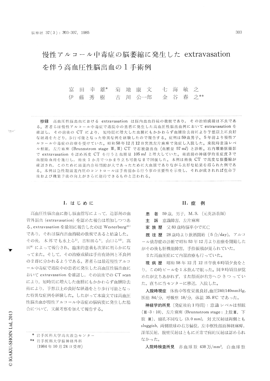
抄録 高血圧性脳出血におけるextravasationは脳内出血持続の徴候であり,その治療成績は不良である。著者らは慢性アルコール中毒症で通院中の患者に発生した高血圧性脳出血例においてextravasationを確認し,その前後のCTにより,短時間に増大した血腫にもかかわらず血腫除去術により予想以上に良好な経過をたどり,歩行可能となった特異な例を経験したので報告する。症例は59歳男子。5年前より慢性アルコール中毒症の治療を受けていた。昭和58年12月12日突然左片麻痺で発症し入院した。来院時意識レベル傾眠,左片麻痺(Brunnstrom stageIII,III) CTで右被殻出血(血腫量57ml)と診断。右内頸動脈撮影でextravasationを認め再度CTを行うと血腫量105 mlと増大していた。術直前の神経学的重症度3で血腫除血術を施行し,術後1か月でつかまり立ち可能なまで同復した。本例は術後CTで高度な脳萎縮が確認され,このために頭蓋内余裕間隙が大であったために大血腫でありながら良好な結果を得られた例である。本例は急性期頭蓋内圧のコントロールは手術前から行う事の重要性を示唆し,それが成されれば生命予後および機能予後の向上がさらに期待できるものと思われる。
It is said that the prognosis is generally unfa-vorable in patients with hypertensive intracerebral hemorrhage showed extravasation on angiogram. Recently, we experienced a case whose prognosis was evenful after the removal of hematoma. So we reported this case and discussed between chronic alcoholism and intracerebral hemorrhage accompanied with extravasation on angiogram in this paper.
A 59-year-old male was transferred to our emer-gency center because of right hemiplegia and mild clouding of consciousness at around nine in the evening on December 12, 1983. At the time of admission, his neurological state was classified into grade III, exhibiting the right putaminal hemorrhage and pyramidal destruction type of hematoma by CT scan with 58 ml of hematoma volume. The right carotid angiography was im-mediately performed and confirmed the hematoma being of lateral type. At that time, extravasationproximal to the lateral lenticulostriate artery was noted. Repeated CT scan revealed the enlarged hematoma (105 ml) accompanying with ventricular hemorrhage. At the completion of these exami-nations, the neurological grade was III. The hematoma was surgically removed after 4 hours following the onset of cerebral hemorrhage. His postoperative course was very favorable. Although acute hydrocephalus appeared later on, it was cured by ventricular drainage. The patient become pos-sible to walk with a helper by 1 month after operation. CT scan obtained 1 month after ope-ration revealed a remarkable brain atrophy, which was probably derived from chronic alcoholism. It is seemed that the intracranial space caused by this brain atrophy may relieve the increased of intracranial pressure in the acute stage, which ultimately resulted in a controlled intracranial pressure required for the acute stage in the course of nature. Thus, even in such cases in danger as showing extravasation, a strict control of intra-cranial pressure followed by craniotomy, removal of hematoma and adequate treatment of hemor-rhagic source may lead to an increased number of patients to be saved.

Copyright © 1985, Igaku-Shoin Ltd. All rights reserved.


