Japanese
English
- 有料閲覧
- Abstract 文献概要
- 1ページ目 Look Inside
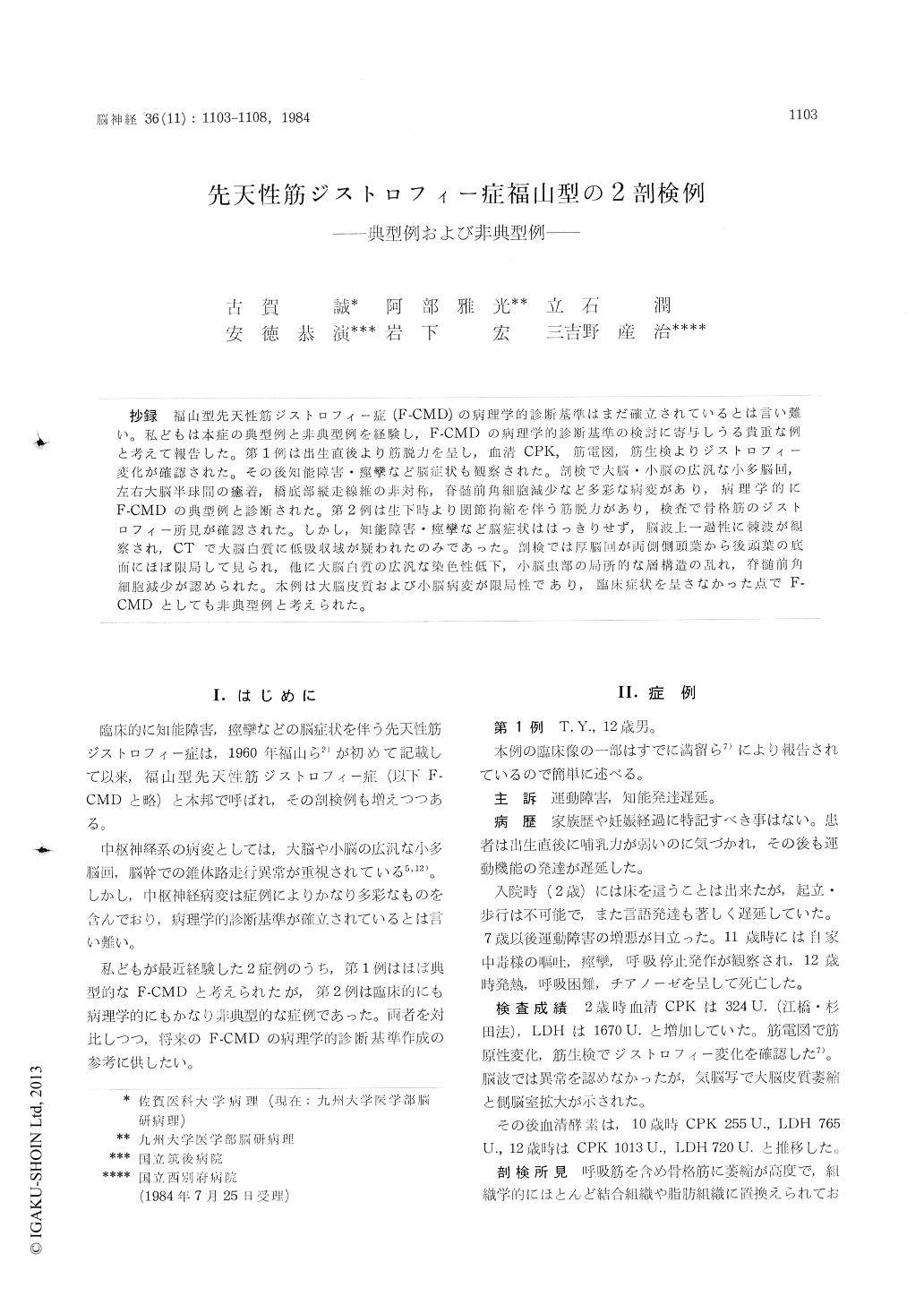
抄録 福山型先天性筋ジストロフィー症(F-CMD)の病理学的診断基準はまだ確立されているとは言い難い。私どもは本症の典型例と非典型例を経験し,F-CMDの病理学的診断基準の検討に寄与しうる貴重な例と考えて報告した。第1例は出生直後より筋脱力を呈し,血清CPK,筋電図,筋生検よりジストロフィー変化が確認された。その後知能障害・痙攣など脳症状も観察された。剖検で大脳・小脳の広汎な小多脳回,左右大脳半球間の癒着,橋底部縦走線維の非対称,脊髄前角細胞減少など多彩な病変があり,病理学的にF-CMDの典型例と診断された。第2例は生下時より関節拘縮を伴う筋脱力があり,検査で骨格筋のジストロフィー所見が確認された。しかし,知能障害・痙攣など脳症状ははっきりせず,脳波上一過性に棘波が観察され,CTで大脳白質に低吸収域が疑われたのみであった。剖検では厚脳回が両側側頭葉から後頭葉の底面にほぼ限局して見られ,他に大脳白質の広汎な染色性低下,小脳虫部の局所的な層構造の乱れ,脊髄前角細胞減少が認められた。木例は大脳皮質および小脳病変が限局性であり,臨床症状を呈さなかった点でF—CMDとしても非典型例と考えられた。
Two autopsy cases of congenital muscular dys-trophy of Fukuyarna type (F-CMD) were described. The first case was diagnosed clinically and patho-logically as its typical case. Neither his family history nor the history of his prenatal period were contributory. He had suffered from muscle weakness and atrophy since his birth. Serum CPK was markedly elevated. EMG and muscle biopsy proved dystrophic changes of the skeletal muscles. In addition, he manifested mental retardation and attacks of convulsion. EEG failed to elicit remark-able changes, but PEG represented ventricular dilatation. He died of respiratory insufficiency at age 12.
His postmortem examination showed variegated anomalies in the nervous system. Extensive mic-ropolygyria was present in the cerebrum and cere-bellum accompanied by adhesions between the bilateral cerebral hemispheres. Assymmetry of the longitudinal fibers was pointed out in the pontine base. Anterior horn cells were atrophic and mo-derately depopulated.
On the other hand, the second patient was an atypical F-CMD case in symptoms, signs and pa-thology. His grand-mothers on both father's and mother's sides were first cousins. His three sib-lings showed no similar disorders. His mother developed slight gestational toxicosis in the sixth and seventh months of pregnancy.
His muscle weakness, contracture of the bilateral hip-joints and clubfoot had been observed since his birth. Physical and neurological examinations at age 6 showed deformity of the skull, myopathic face, macroglossia, high-arched palate, pigeon chest, scoliosis of the thoracic spine. In addition, generalized muscular atrophy, hypotonia and are-flexia were recognized. Pseudohypertrophy of the muscles was absent. Sensation was intact to all modalities. Serum CPK and LDH were moderately increased. EMG and muscle biopsy resulted in dystrophic changes of the skeletal muscles.
No definite brain s3noptcins were elicited. Men-tal retardation was not remarkable and his IQ was between 71 and 92. Thcugh he had no episo-des of convulsion, diffuse epileptic pattern was observed temporarily in EEG. Computerized to-mography showed low density area bilaterally around the lateral ventricles. He died of respira-tory insufficiency at age 15.
His postmortem investigation revealed localized pachygyric micropolygyria, which was confined to the bilateral temporal and occipital bases. Myelin stains showed extensive pallor in the cerebral white matter. Focal dysarrangement of the archi-tecture was found also in the cerebellar vermis. Anterior horn cells in the spinal cord were atro-phic and decreased in number. Neither inflamma-tion nor fibrous thickening were seen in the me-ninges.
In the majority of the reported cases of F-CMD, the nervous system lesions were extensive includ-ing outstanding and widespread micropolygyria in the cerebrum and cerebellum like our first case. Only a few reports described focal involvement of the cerebral and cerebellar cortices similar to our second case. Present cases suggest that there may be a wide range of variation in localization and extent of CNS lesions in F-CMD, including forme fruste.

Copyright © 1984, Igaku-Shoin Ltd. All rights reserved.


