Japanese
English
- 有料閲覧
- Abstract 文献概要
- 1ページ目 Look Inside
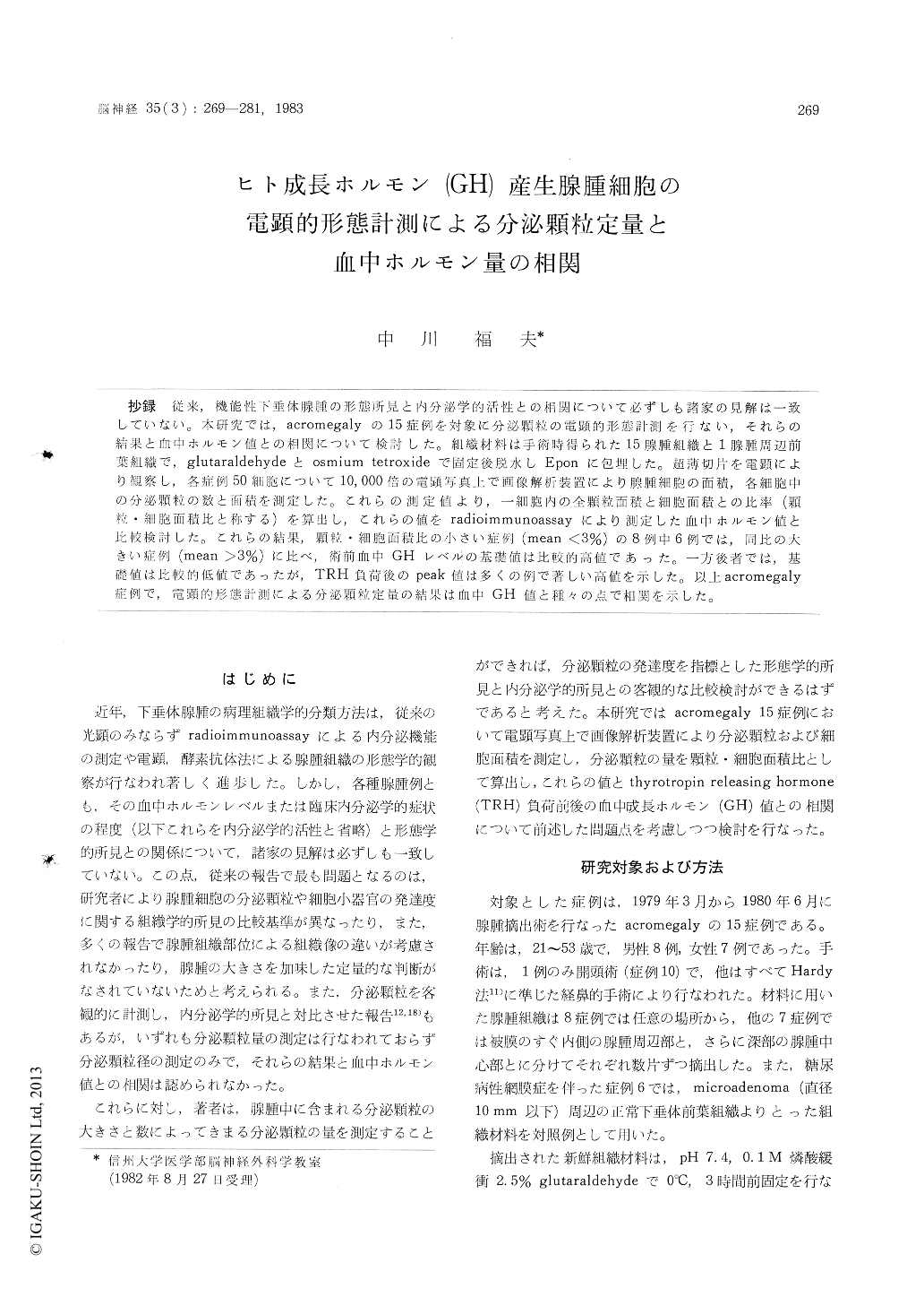
抄録 従来,機能性下垂体腺腫の形態所見と内分泌学的活性との相関について必ずしも諸家の見解は一致していない。本研究では,acromegalyの15症例を対象に分泌顆粒の電顕的形態計測を行ない,それらの結果と血中ホルモン値との相関について検討した。組織材料は手術時得られた15腺腫組織と1腺腫周辺前葉組織で,glutaraldehydeとosmium tetroxideで固定後脱水しEponに包埋した。超薄切片を電顕により観察し,各症例50細胞について10,000倍の電顕写真上で画像解析装置により腺腫細胞の面積,各細胞中の分泌顆粒の数と面積を測定した。これらの測定値より,一細胞内の全顆粒面積と細胞面積との比率(顆粒・細胞面積比と称する)を算出し,これらの値をradioilnmunoassayにより測定した血中ホルモン値と比較検討した。これらの結果,顆粒・細胞面積比の小さい症例(mean<3%)の8例中6例では,同比の大きい症例(mean>3%)に比べ,術前血中GHレベルの基礎値は比較的高値であった。一方後者では,基礎値は比較的低値であったが,TRH負荷後のpeak値は多くの例で著しい高値を示した。以上acromegaly症例で,電顕的形態計測による分泌顆粒定量の結果は血中GH値と種々の点で相関を示した。
In studies of functioning pituitary adenomas, different findings have been reported concerning the correlation between the amount of secretory granules as determined by light or electron microscopic observation and endocrinological findings. In the present study, the author exam-ined the correlation by means of electron microscopic morphometry.
Sixteen specimens were obtained at the time of surgery from 15 acromegalic patients : 15 adenoma tissues and 1 normal control anterior pituitary tissue from a patient with acromegaly accompanied by diabetic retinopathy. All the specimens were fixed in buffered glutaraldehyde and osmium tetroxide, dehydrated and embedded in epon. Thick sections were stained with toluidine blue and were observed by light microscopy to obtain a gross impression of the histology and the amount of secretory granules in the adenoma cells.
For the morphometric study of secretory gran-ules, thin sections were stained with lead citrateand uranyl acetate, examined by electron micro-scopy and photographed. On electron photomicro-graphs, magnified 10, 000×, both numbers and areas of secretory granules in a cell and the area of the cell itself were measured for 50 cells from each specimen by an image-analyser (Digigramer-G, manufactured by Mutoh Industrial Company, Tokyo). From the data, the area ratios of granules to the cells to which they belonged (G/C ratios) were calculated. The plasma growth hormone (GH) and prolactin (PRL) levels from correspond-ing patients were measured, and these results were compared with the morphometric results.
The results obtained were as follows :
1) All the cases were divided into two groups (Groups I and II) by mean values of G/C ratios. The seven cases in Group I showed mean G/C ratios over 3%, and the 8 cases of Group II under 3% (Table 1, Fig. 4).
2) Preoperative basal serum GH levels were relatively higher in 6 out of the 8 cases of Group II than in the 7 cases of Group I. The other 2 cases in Group II showed relatively lower serum GH levels. The area of most secretory granules in these 2 cases was small- er than 0. 02 /tm' (Fig. 4, 5, 7).
3) In the 7 cases of Group I, preoperative serum GH levels before the administration of TR1-I were relatively low but, after the administra- tion of TRH, remarkably high (Fig. 4, 7).
4) In the 7 cases of Group I, distribution of the number of cells as classified according to the size of granules resembled that in normal control somatotrophs (Fig. 4).
5) Two cases with high prolactinemia belonged to Group II (Table 1).
From the results, it is proposed that in acro-megaly some correlation exists between the results obtained by electron microscopic morphometry of secretory granules from adenoma cells and the serum hormonal levels.

Copyright © 1983, Igaku-Shoin Ltd. All rights reserved.


