Japanese
English
- 有料閲覧
- Abstract 文献概要
- 1ページ目 Look Inside
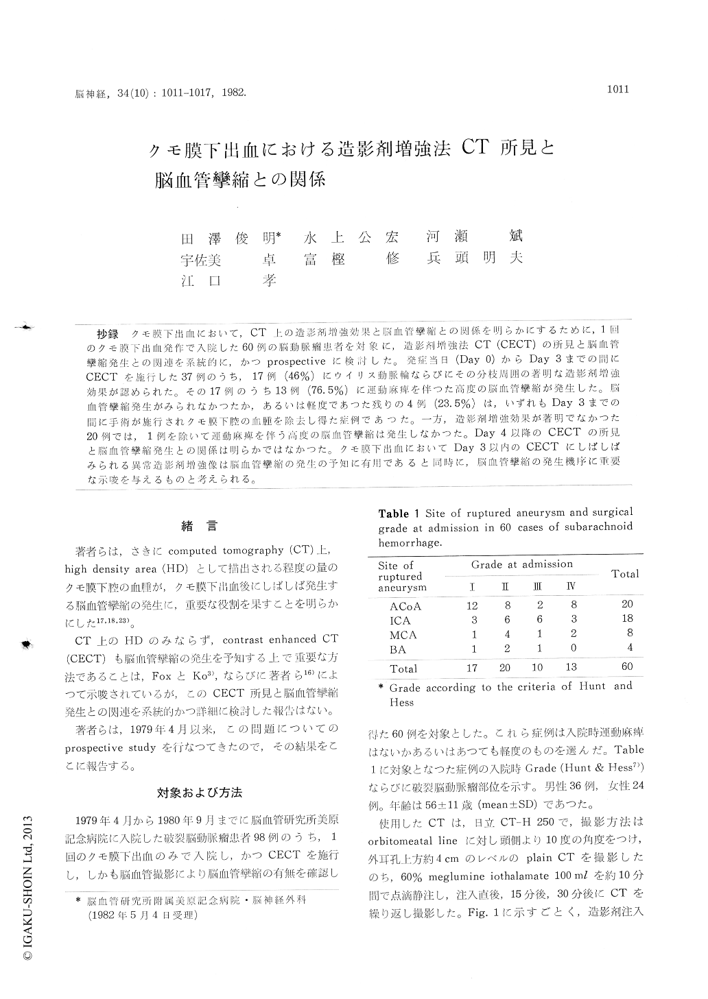
抄録 クモ膜下出血において,CT上の造影剤増強効果と脳血管攣縮との関係を明らかにするために,1回のクモ膜下出血発作で入院した60例の脳動脈瘤患者を対象に,造影剤増強法CT (CECT)の所見と脳血管攣縮発生との関連を系統的に,かつprospectiveに検討した。発症当日(Day 0)からDay 3までの間にCECTを施行した37例のうち,17例(46%)にウイリス動脈輪ならびにその分枝周囲の著明な造影剤増強効果が認められた。その17例のうち13例(76.5%)に運動麻痺を伴つた高度の脳血管攣縮が発生した。脳血管攣縮発生がみられなかつたか,あるいは軽度であつた残りの4例(23.5%)は,いずれもDay 3までの間に手術が施行されクモ膜下腔の血側腫を除去し得たり症例であつた。一方,造影剤増強効果が著明でなかつた20例では,1例を除いて運動麻痺を伴う高度の脳血管攣縮は発生しなかつた。Day 4以降のCECTの所見と脳血管攣縮発生との関係は明らかではなかつた。クモ膜下出血においてDay 3以内のCECTにしばしばみられる異常造影剤増強像は脳血管攣縮の発生の予知に有用であると同時に,脳血管攣縮の発生機序に重要な示唆を与えるものと考えられる。
We have often experienced that delayed cere-bral vasospasm is preceded by prominent increase in density in the region of the circle of Willis and its branches on contrast enhanced CT (CECT). This prospective study was undertaken in order to know the relationship between this phenomenon on CECT and cerebral vasospasm in patients with subarachnoid hemorrhage.
Sixty patients with a single rupture of an aneurysm were subjected to study. Contrast enhanced CT was performed by intravenous infusion of 100 ml of 60% meglumine iothalamate in 10 minutes. Post-contrast CT scans were repeated serially just after infusion, 15 minutes and 30 minutes later. Prominent increase in density in the region of the circle of Willis and its bran-ches 30 minutes after infusion was considered as remarkable enhancement. Angiography was per-formed on admission and was repeated betweenday 4 and day 14 (the day of subarachnoid hemor-rhage was day 0) in order to check the develop-ment of vasospasm in each case. Only motor weakness associated with angiographic vasospasm was considered as ischemic symptom due to vaso-spasm in this study. Twenty-seven patients un-derwent the operation of aneurysm within day 3 and 28 patients underwent the operation between day 4 and day 30. Removal of subarachnoid clot was performed as much as possible during the operation.
In 17 (46%) out of 37 patients who under went CECT within day 3, the contrast enhan-cement was remarkable. Transient or permanent symptomatic vasospasm occured in 13 (76.5%) out of these 17 patients and the remaining 4 patients who underwent the operation with successful removal of subarachnoid clot within day 3 did not develop symptomatic vasospasm. Eight (67%) out of 12 patients operated within day 3, in whom post-operative CT showed incomplete removal of subarachnoid clot, developed transient or perma-nent symptomatic vasospasm. In only one ( 5 %) out of 20 patients without remarkable enhance-ment, transient symptomatic vasospasm occured. The abnormal contrast enhancement in the region of the circle of Willis and its branches within day 3 was closely related to the subsequent occur-rence of vasospasm.
Contrast enhanced CT was performed in 41 patients after day 3. There was no patient with remarkable enhancement on CECT. There was no relationship between the findings on CECT after day 3 and the occurrence of vasospasm.
In order to elucidate the mechanism of remarka-ble enhancement, we have calculated iodine content in circulating blood and estimated iodine content in the region of the circle of Willis by analyzing the quantitative aspects of the CT scans both before and after injection of contrast medium according to Gado's study. The rate of decrease of iodine content in the region of the circle of Willis was apparently lower than that in the circulating blood. This abnormal increase in density in the region of the circle of Willis after contrast infusion is, therefore, apparent leakage of contrast medium from the vessels. We consider that this remarkable enhancement of subarachnoid space indicates the increase of permeability of small vessels especially of the venular side due to aseptic meningitis caused by subarachnoid clot.
We believe that CECT within day 3 allows prediction of that patient destined for vasospasm and early removal of subarachnoid clot within day 3 may minimize the future development of vasospasm.

Copyright © 1982, Igaku-Shoin Ltd. All rights reserved.


