Japanese
English
- 有料閲覧
- Abstract 文献概要
- 1ページ目 Look Inside
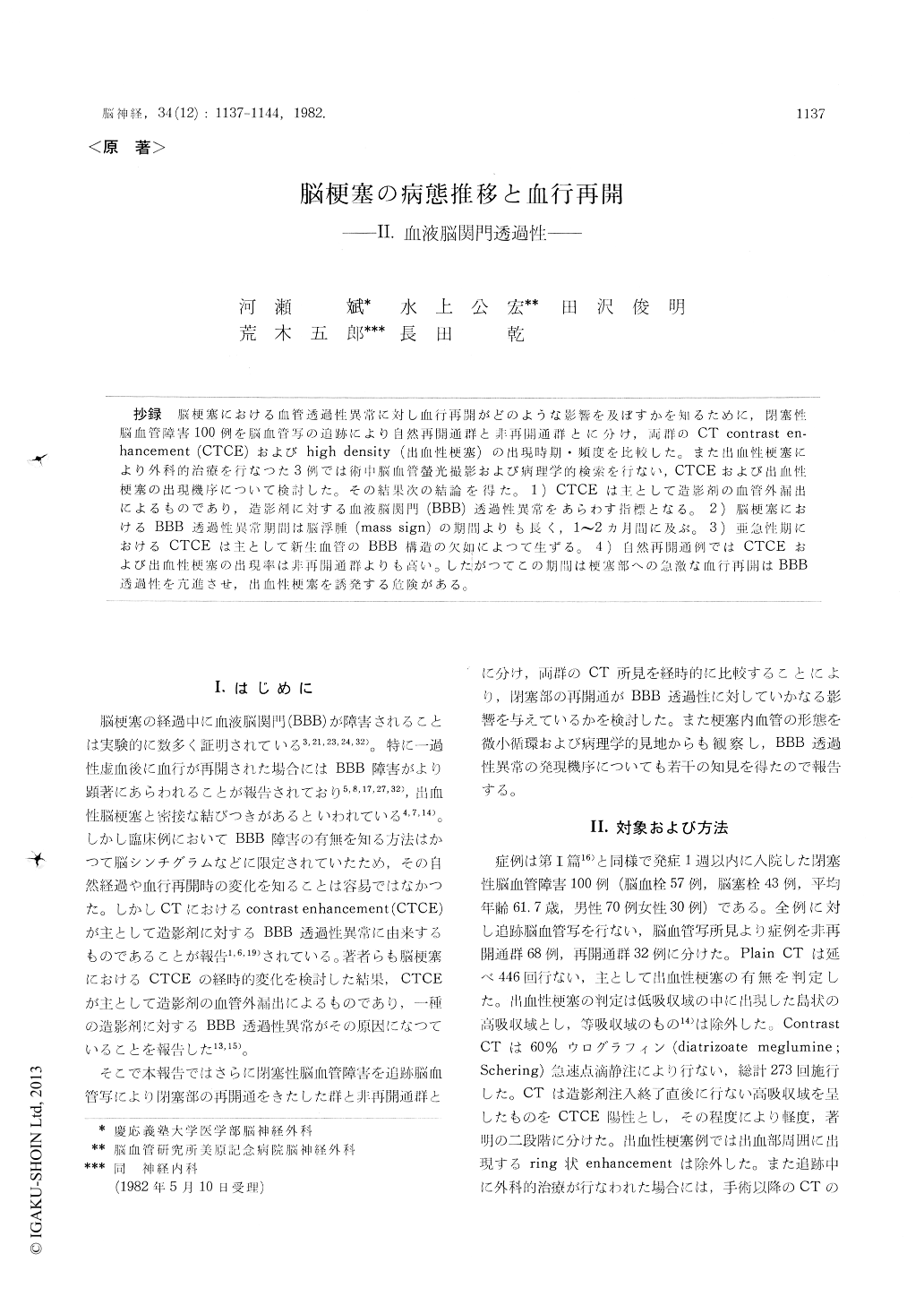
抄録 脳梗塞における血管透過性異常に対し血行再開がどのような影響を及ぼすかを知るために,閉塞性脳血管障害100例を脳血管写の追跡により自然再開通群と非再開通群とに分け,両群のCT contrast en—hancement (CTCE)およびhigh density (出血性梗塞)の出現時期・頻度を比較した。また出血性梗塞により外科的治療を行なつた3例では術中脳血管螢光撮影および病理学的検索を行ない,CTCEおよび出血性梗塞の出現機序について検討した。その結果次の結論を得た。1) CTCEは主として造影剤の血管外漏出によるものであり,造影剤に対する血液脳関門(BBB)透過性異常をあらわす指標となる。2)脳梗塞における BBB透過性異常期間は脳浮腫(mass sign)の期間よりも長く,1〜2カ月間に及ぶ。3)亜急性期におけるCTCEは主として新生血管のBBB構造の欠如によつて生ずる。4)自然再開通例ではCTCEおよび出血性梗塞の出現率は非再開通群よりも高い。したがつてこの期間は梗塞部への急激な血行再開はBBB透過性を亢進させ,出血性梗塞を誘発する危険がある。
One hundred patients with occlusive cerebro-vascular diseases were studied to know the sequential change of blood-brain barrier (BBI3) permeability in ischemic stroke. The disruption of BBB was represented as contrast enhancement on computerized tomography (CTCE) and a manifestation of high density focus in the in-farcted zone (hemorrhagic infarction). A total of 446 studies of plain CT and 273 studies of contrastCT were performed. Angiography was followed in all patients and they were allocated to two groups according to the findings on angiograms : 68 patients having any change of occlusive lesion (the group of no-recanalization) and 32 patients showing reopening of occluded vessels (the group of recanalization). Fluorescein cerebral angio-graphy (FCA) and pathological examination were done in 3 patients who underwent surgery due to hemorrhagic infarction occurred in subacute stage of infarction.
In most cases, CTCE became manifest 7 days or more following stroke, and had the peak in-cidence in the third week, and disappeared between one and two month. In the no-recanalized group, CTCE did not occurred during the edema stage (2 to 8 days). Hemorrhagic infarction was rare in no-recanalized group (4%). Whereas in the recanalized group, CTCE appeared even in the acute stage, only when contrast CT was performed immediately after recanalization. CTCE was prominent in all cases during one month of stroke, and hemorrhagic infarction was present in 15 cases (47%). In those cases, high density focus appeared also in the subacute stage (one week to one month) as well as in the acute stage (within one week), and marked CTCE usually preceded the appearance of high density focus. Extravasation of fluorescein dye was shown in those CTCE zone. Pathological specimen demon-strated findings similar to those seen in the resolution stage of infarction. Numerous imma-tured capillaries, without basement membrane, extended from cortex to subcortex, and extra-vasated erythrocytes were found in the perivas-cular spaces.
It is conceivable, therefore, that the reopening of occluded vessels accelerates the permeability of disrupted BBB, which is resulted as marked CTCE and hemorrhagic infarction. Disruption of BBB may be caused not only by ischemic damage of surviving vessels but also by newly formed vessels which are not provided with a BBB in the subacute stage. Low perfusion during ischemia and cerebral edema is considered to supress the BBB permeability.

Copyright © 1982, Igaku-Shoin Ltd. All rights reserved.


