Japanese
English
- 有料閲覧
PPV購入案内
1,320円 (1,200円+税10%) こちらは、151ページから158ページの文献です。購入には医書.jp本会員登録が必要です。
会員登録完了後に改めて上記「購入サイトへ」を選択してください。
または「購入サイトへ」選択後に「購入ログイン」、「新規会員登録」の順に進んでください。
会員登録について詳しくはをご確認ください。
- Abstract 文献概要
- 1ページ目 Look Inside
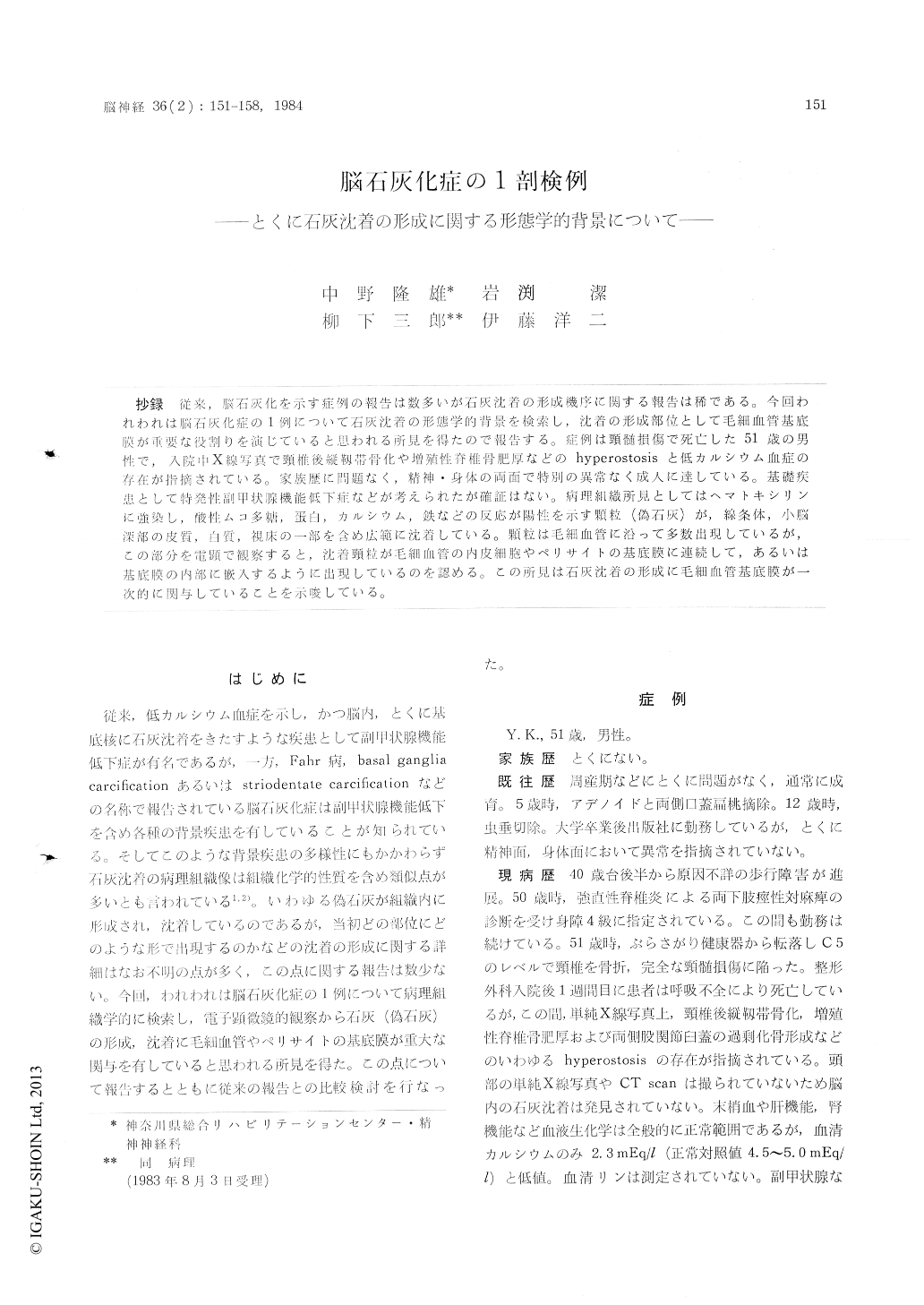
抄録 従来,脳石灰化を示す症例の報告は数多いが石灰沈着の形成機序に関する報告は稀である。今回われわれは脳石灰化症の1例について石灰沈着の形態学的背景を検索し,沈着の形成部位として毛細血管基底膜が重要な役割りを演じていると思われる所見を得たので報告する。症例は頸髄損傷で死亡した51歳の男性で,入院中X線写真で頸椎後縦靱帯骨化や増殖性脊椎骨肥厚などのhyperostosisと低カルシウム血症の存在が指摘されている。家族歴に問題なく,精神・身体の両面で特別の異常なく成人に達している。基礎疾患として特発性副甲状腺機能低下症などが考えられたが確証はない。病理組織所見としてはヘマトキシリンに強染し,酸性ムコ多糖,蛋白,カルシウム,鉄などの反応が陽性を示す願粒(偽石灰)が,線条体,小脳深部の皮質,白質,視床の一部を含め広範に沈着している。顆粒は毛細血管に沿って多数出現しているが,この部分を電顕で観察ずると,沈着頸粒が毛細血管の内皮細胞やペリサイトの基底膜に連続して,あるいは基底膜の内部に嵌入するように出現しているのを認める。この所見は石灰沈着の形成に毛細血管基底膜が—次的に関与していることを示唆している。
The case was a 51 years old male who died of cervical spinal cord injury. On admission, X-ray disclosed distinct hyperostosis such as ossifi-cation of posterior longitudinal ligament, ankylo-sing spondilitis and callus luxurians of bilateral hip joints.
He had no familial history. He normally deve-loped into adult without any mental and neurolo-gical abnormalities. In the latter half of the fifth decade, he developed progressive spastic diplegiaof his legs. Laboratory studies revealed evident hypocalcaemia (2.3 mEq/l). Hormonal examination of the parathyroid gland was not performed.
Postmortem examination of the brain (1,400 g) disclosed widely spreading numerous spherical deposits of various sizes stained deeply with he-matoxilin. These deposits were positive with PAS, colloidal iron, prussian blue and Kossa's Method for calcium etc. as shown in Table 1.
According to their histochemical properties, these deposits were considered to consist of both acid mucopolysaccharides and proteins to which calcium and iron have been bound later. These deposits were predominantly observed in the basal ganglia, dorso-lateral portion of the thalamus and the depth of the cerebellum. The dentate nucleus was mostly spared. To the lesser degree, these deposits were also seen around the capillaries andsubadventitial space of the small vessels in the cerebral cortex, cerebral white matter, capsula interna and red nucleus. These deposits were sho-wn to be adjacent to the capillary walls micro-scopically.
Electron microscopy disclosed that many elect-ron dense spherical bodies surrounding capillary were related to the basement membrane of the endothelium or pericyte. Smaller ones seemed to be encased by the basement membranes. Both neu-ronal and glial cells were well preserved and see-med to have no correlation with the formation of the deposits. It was considered that the basement membrane of the capillary might be the initial site of the formation and developement of the deposits, followed by calcium or iron binding.
Copyright © 1984, Igaku-Shoin Ltd. All rights reserved.