Japanese
English
- 有料閲覧
- Abstract 文献概要
- 1ページ目 Look Inside
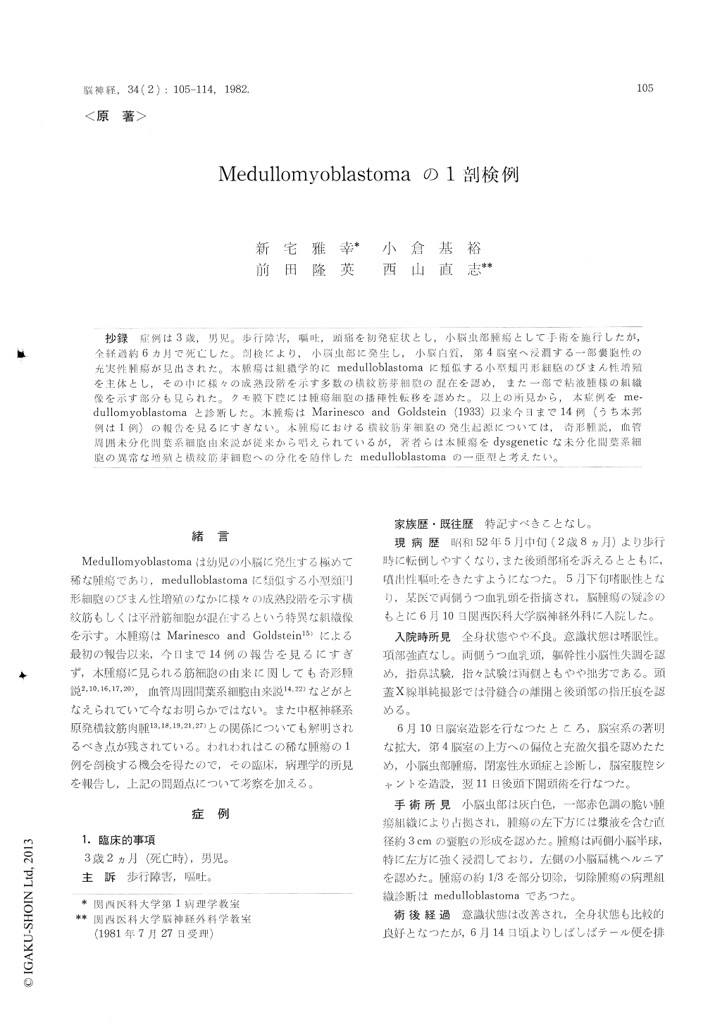
抄録 症例は3歳,男児。歩行障害,嘔吐,頭痛を初発症状とし,小脳虫部腫瘍として手術を施行したが,全経過約6ヵ月で死亡した。剖検により,小脳虫部に発生し,小脳白質,第4脳室へ浸潤する一部嚢胞性の充実性腫瘍が見出された。本腫瘍は組織学的にmedulloblastomaに類似する小型類円形細胞のびまん性増殖を主体とし,その中に様々の成熟段階を示す多数の横紋筋芽細胞の混在を認め,また一部で粘液腫様の組織像を示す部分も見られた。クモ膜下腔には腫瘍細胞の播種性転移を認めた。以上の所見から,本症例をme—dullomyoblastomaと診断した。本腫瘍はMarinesco and Goldstein (1933)以来今日まで14例(うち本邦例は1例)の報告を見るにすぎない。本腫瘍における横紋筋穿細胞の発生起源については,奇形腫説,血管周囲未分化間葉系細胞由来説が従来から唱えられているが,著者らは本腫瘍をdysgeneticな未分化間葉系細胞の異常な増殖と横紋筋芽細胞への分化を随伴したmedulloblastomaの一亜型と考えたい。
An autopsy case of medullomyoblastoma was presented. The patient was admitted to the hospital at the age of 2 years and 8 months, because of an unsteady gait and projectile vomiting lasting for a month. Ventriculography at the admission dis-closed a filling defect in the fourth ventricle, so the tumor arising from the cerebellar vermis was suspected. Posterior fossa craniotomy was performed and the tumor in the cerebellar vermis was partially resected. Postoperatively, despite of the irradiation therapy, the general condition of the patient was gradually deteriorated, and the patient died at the age of 3 years and 2 months, 6 months after the onset of the clinical symptoms.
Autopsy revealed obstructive hydrocephalus, and solid tumor in yellowish grey color in the cerebel-lar vermis, which showed diffuse infiltration to the neighbouring cerebellar white matter and to the subarachnoid cavity at the level of the lumbar cord. The tumor was partly cystic, and the tumor protruding into the fourth ventricle showed the gelatinous cut surface.
Histologically, the tumor was mainly composed of densely proliferating small round cells with hyperchromatic nuclei. This finding resembles closely medulloblastoma but, in addition, includes unequivocal cross-striated muscle fibers. Neither rosette of Homer-Wright type nor perivascular pseudorosette were detected. There was no evidence suggesting the differentiation to the neuronal or glial cells. Rhabdomyoblastic cells in the varying stage of differentiation, which were intimately in-termingled with these small round cells, were found in the relatively localized area in the vermis. Most of these rhabdomyoblastic cells showed remarkable pleomorphism and distinct cross-striation in their cytoplasm. Some of these cells formed mature muscle fibers. In the metastatic foci disseminated in the subarachnoidal cavity, no rhabdomyoblastic cell was detected. In addition, the myxomatous area was found in the tumor, corresponding to the tumor mass whose cut surface showing the gela-inous appearance. These areas were composed of sparsely proliferating undifferentiated mesenchymal cells with abundant myxoid stroma and scattered muscle fibers.
From the above findings, this tumor was diag-nosed as very rare "medullomyoblastoma" (Mari-nesco and Goldstein), being corresponding to the fifteenth case reported in the world literature. The clinicopathological features of this rare tumor were reviewed in comparison with those of primaryrhabdomyosarcoma of the central nervous system. Besides, the origin of rhabdomyoblastic cells in medullomyoblastoma were discussed. The present authors considered rhabdomyoblastic cells to be originated from perivascular undifferentiated mesen-chymal cells, and this tumor as a variant of medul-loblastoma accompanied by the proliferation and aberrant differentiation of dysgenetic mesenchymal cells.

Copyright © 1982, Igaku-Shoin Ltd. All rights reserved.


