Japanese
English
- 有料閲覧
- Abstract 文献概要
- 1ページ目 Look Inside
抄録 Sturge-Weber症候群の脳血管撮影において,従来ほとんど異常を認めないのが普通と考えられてきたが,最近Venous angiomaをはじめ脳静脈系の異常が指摘されてきた。我々は本症候群の脳血管撮影で両側にまたがる広範な脳静脈・静脈洞の異常を示し,更に脳静脈還流異常を呈した症例を経験したのでここに報告する。本症例は2歳男児で顔面の正中よりやや左側と頭皮や四肢にも血管性母斑を持ち,神経症状として痙攣発作と左片麻痺を認めた。脳波で右頭頂側頭部にspikeが出現し頭部単純撮影で異常石灰化像は認められなかつたが,CT scanにて右頭頂後頭部に石灰化像がみられ,さらに同部分に造影剤による増強効果を認め,主たる頭蓋内病変が右頭頂後頭葉に存在するSturge-Weber症候群と診断した。また脳血管撮影で胎生期に存在するprimitive straight sinusの遺残をはじめ脳表面から脳深部にわたり,しかも両側にまたがる広範な脳静脈・静脈洞の異常および脳静賑還流異常を認めた。我々は本症例にみられたような広範な血管変化はSturge-Weber症候群の原因として考えられているplexiform layerの発達異常に加え,さらにその後の静脈洞および皮質静脈などの脳静脈発達途上での異常を伴つたものと考える。
A case of Sturge-Weber syndrome with marked abnormalities in the cerebral venous system was reported.
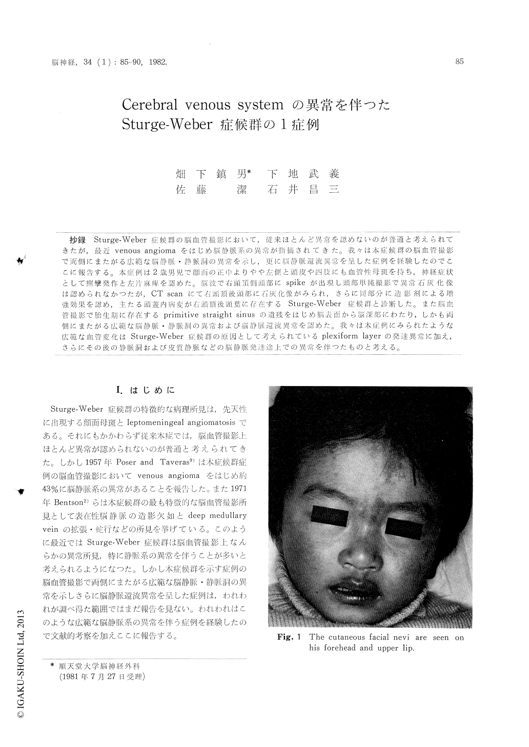
The patient was a 2-year old boy who was admit-ted to the Department of Neurosurgery with the chief complaints of left hemiparesis and left focal seizures. He had vascular nevi on the forehead and upper lip of his face, scalp, right forearm and thigh (Fig. 1).
Neurological examination of admission revealed left hemiparesis.
Plain skull films indicated no intracranial calcifi-cation.
EEG showed paroxysmal focus in the right parieto-temporal area.
Plain CT scan showed calcium deposits in the right parietooccipital area and contrast enhance-ment occurred around the areas of calcification (Fig. 2).
Venous phases of bilateral CAGs showed abnor-malities of the cortical veins and sinuses and abnormal drainage from the cerebral cortex to the deep veins.
It also demonstrated persistence of the primitive straight sinus (Fig. 3, 4).
From the neurological and neuroradiological findings, this case was diagnosed as the Sturge-Weber syndrome with marked abnormalities in the cerebral venous system.
These abnormal findings of veins and sinuses seemed to be brought about by development abnor-malities of veins and sinuses which continuously occurred following Streeter's primordial plexus, which has been considered to be a cause of the Sturge-Weber syndrome.
Copyright © 1982, Igaku-Shoin Ltd. All rights reserved.