Japanese
English
- 有料閲覧
- Abstract 文献概要
- 1ページ目 Look Inside
I.はじめに
Gliomaの放射線療法は術後補助療法として不可欠である。今日超高圧X線(60Co,Linac)や超高圧電子線(Betatron)のような低LET線源による照射が一般に用いられているが,その治療効果を可能な限り高めるために,さまざまの工夫が各方面からなされている1,13,18)。その一つの方法として,glioma細胞を放射線感受性の高いG2—M移行期に同調蓄積させ,放射線照射を行う方法は理論的にも意義深いものがあり,初期導入療法として良い成績をおさめている19,22)。
G2—M期への同調剤(synchronizer)の細胞静止作用を生長解析パラメータ14)を用いて解析することは重要であるがflow cytometry (FCM)は,この目的にかなう価値の高い検査法である12,28)。現在までその解析には腫瘍細胞を全集団として分析していたが,腫瘍細胞の正確な生長解析を行うには同調された同一細胞周期を進行する細胞だけを集めることができれば理想的である。
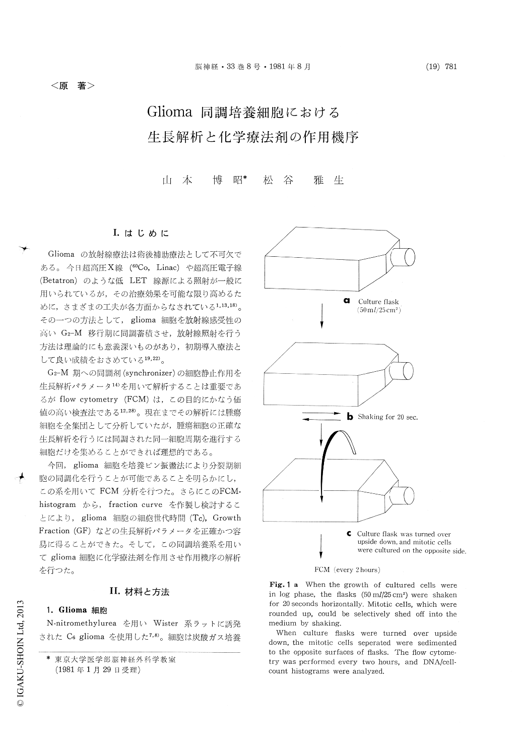
Mitotic cells of cultured C6 glioma, which were rounded up and could easily be shed off, were selected by shaking the culture flasks. The culture flasks were then turned over upside down, and mitotic cells were cultured on the oposite surfaces of the flasks. The synchronized cultured cells proceeded through the succeding new cell cycle. The flow cytometric analysis was performed in every two hours during 30 hours after the separa-tion. DNA distribution histograms were obtained, and the ratio of the number of cells in each cell cycle phase vs. total cell population was calculated by Fried's method.
The fraction curve of cells in each cell cycle phase -G1, S, and G2+M- was thus determined as a function of time, corresponding to fG1(t), fs (t), and fG2+M (t).
The cell kinetic parameters of C6 glioma, Tc (cell cycle time) and GF (Growth Fraction) were calculated. Tc was at first defined as summation of TG1, TS, and TG2+M (Method 1). It was also defined as the period between the times of the initiation of mitotic cell synchrony and maximum resynchrony of cells in mitotic phase (Method 2).
Tc calculated by Method 1 and 2 was 19.8 and 20.8 hours respectively. GF was defined as maxi-mum altering range of value of fG1(t), and was calculated as 0.8.
Mode of action of chemotherapeutic agents (vincristine, ACNU, and VM 26) was also investi-gated using this synchronized C6 glioma. Vincri-stine acted as a G2-M phase blocker at the con-centration of 0.005μg/ml. But at the higher concentration (more than 0.01μg/ml), cytostatic effect at middle S phase was detected on the S fraction curve. ACNU inhibited the cell traverse at the middle S phase, and the slow proceeding of synchronized cell population through late S phase to G2-M phase was determined on the S fraction curve (fS (t)). VM 26, at the concentration of 0.01-1μg/ml, specifically acted as a G2-M phase blocker without inhibition of S phase traverse.
By the aid of flow cytometric analysis of various chemotherapeutic agnnts, the therapeutic regimen for brain tumor chemotherapy can be rationally established and the improvement of clinical thera-peutic results of gliomas will be expected.

Copyright © 1981, Igaku-Shoin Ltd. All rights reserved.


