Japanese
English
- 有料閲覧
- Abstract 文献概要
- 1ページ目 Look Inside
I.はじめに
結核性髄膜炎に遭遇する機会は今日稀となり,したがつてCT scanによる報告も僅かしか見あたらない。著者らは結核性髄膜炎に罹患した幼児例を,その発病の初期より半年以上にわたつてCT scanで追跡し,basaltuberculous meningitisを表現すると思われる興味ある所見を得た。本文ではそのCT像を報告するとともに,従来確診の困難であつた結核性髄膜炎の早期診断におけるCT scanの意義について考察を加えたい。
The authors report a case of tuberculous meningitis in which computerized tomography (CT) demonstrated findings presumably of the basalgranulomatous meningitis.
A 11/2-year-old girl presented with a 3-week history of moodiness, anorexia, fever, progressive drowsiness and slight left hemiparesis. Opening pressure on lumber puncture was 300 mm2O. CSF analysis revealed 176 white blood cells with 70% mononuclear cells, 150 mg/100 ml protein and 60-70/100 ml sugar; tryptophan reaction was positive; fibrinous web was formed. CSF stains and cultures were negative. Tuberculous skin test was positive. A chest roentgenogram showed atelectasis of the left upper lobe. The patient had a history of contact with her father who was suffering from active pulmonary tuberculosis. A tentative di-agnosis of tuberculous meningitis was made. Continuous ventricular drainage was performed for the treatment and antituberculous chemotherapy was instituted resulting in cessation of inflammatory process in subsequent months. Then ventriculo-peritoneal shunt was made. But the patient's condition continued to deteriorate and she became vegetative.
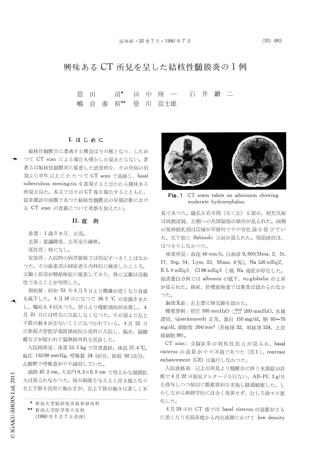
CT on admission showed moderate hydrocephalus. Two days later, CT demonstrated low density area in the right pulvinar and the posterior limb of the internal capsule. The basal cisterns were poorly visualized. With contrast enhancement, abnormal enhancement appeared mainly in the basal cisterns and the neighborhood of the tentorium cerebelli. One month after hospitalization, hydrocephalus progressed and the basal cisterns became isodense and were not identified on plain CT. There were mottled low and high density lesions diffusely ex-tended in the brain parenchyma. The midbrain became low dense. Enhanced CT revealed diffuse enhancement of the basal cisterns. Multiple irregular enhancements were also seen in the brain parenchyma. After six months, severe hydro-cephalus was found. There was localized enhance-ment only in the suprasellar and the right ambient regions.
Similar findings have been described by Enzmann et al. in two patients with coccidioidal meningitis and a patient with tuberculous meningitis, and seem to represent granulomatous meningitis. This could be of great value for the diagnosis of tuber-culous meningitis. The low density seen in the brain parenchyma may probably be due to vascular lesions of the inflammatory process.
Copyright © 1980, Igaku-Shoin Ltd. All rights reserved.