Japanese
English
- 有料閲覧
- Abstract 文献概要
- 1ページ目 Look Inside
I.はじめに
抗結核薬の開発,結核対策の推進,栄養などの生活水準の向上により,近年,結核死亡は確実に減少した.それに比例して,結核性髄膜炎は比較的稀な疾患となったと考えている臨床家が多いと思われる.事実,小児においては比較的稀な疾患となりつつあるが,成人においては疑診の症例も含めると発生率が著明に減少しているとは言えない.結核性髄膜炎は現在でも難治性髄膜炎の一つであり,その予後は他の疾患と比較しても,必ずしも満足のいくものではない.治療薬としてrifampicinが登場してからも,ほとんど改善されていない.抗結核薬の開発に比べて,治療成績が向上しない理由として,本症に対する関心の薄れと共に,結核菌の髄液からの検出率が悪いために治療開始が遅れたり,治療が中途半端で終わってしまっていることが考えられる9,14).
緩解期の抗結核剤投与中止の指標として,CTでのcontrast enhancementの消失が挙げられているが,真に適切な指標であるのかに言及した文献は少ない.本院で経験した結核性髄膜炎の症例を紹介し,緩解期の抗結核薬の投与計画における画像診断の役割について考察したいと思う.
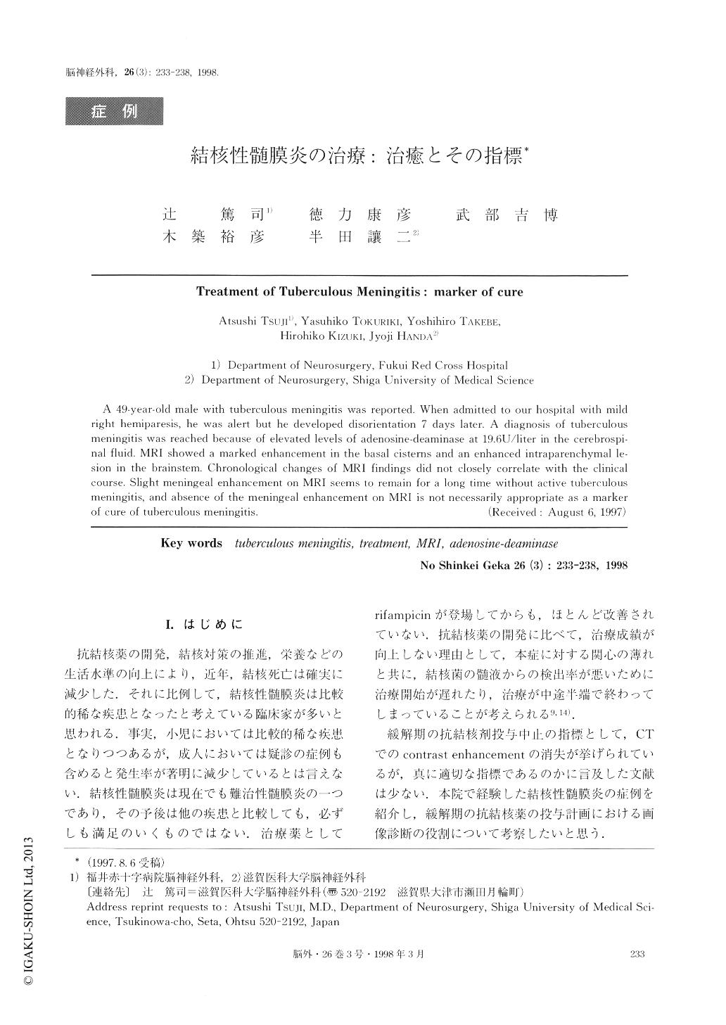
A 49-year-old male with tuberculous meningitis was reported, When admitted to our hospital with mildright hemiparesis, he was alert but he developed disorientation 7 days later, A diagnosis of tuberculousmeningitis was reached because of elevated levels of adenosine-deaminase at 19.6U/liter in the cerebrospi-nal fluid MRI showed a marked enhancement in the basal cisterns and an enhanced intraparenchymal le-sion in the brainstem. Chronological changes of MRI findings did not closely correlate with the clinicalcourse. Slight meningeal enhancement on MRI seems to remain for a long time without active tuberculousmeningltis, and absence of the meningeal enhancement on MRI is not necessarily appropriate as a markerof cure of tuberculous meningitis.

Copyright © 1998, Igaku-Shoin Ltd. All rights reserved.


