Japanese
English
- 有料閲覧
- Abstract 文献概要
- 1ページ目 Look Inside
I.はじめに
中枢神経系のアミノ酸についての知見が集積されるにしたがって,アミノ酸が中枢神経系において多くの重要な役割を果たしていることが明らかとなつてきた。たとえば,興奮の伝達は神経組織において最も根本的な機能であるがGABA, glutamateなどのアミノ酸が,神経伝達物質の1つとして働いていることが知られている10,22)。また,alanine,glutamate,aspartate等,多くのアミノ酸は,嫌気的および好気的糖代謝と密接な関係を有し,そのreservoir poolとして働いている。したがって,脳の機能や代謝の異常により脳内遊離アミノ酸レベルが変動し,またこのアミノ酸レベルの変動が脳の機能や代謝に影響を及ぼすことは十分に考えられることである。
低血糖12)hypercapnea3,25),hypoxya1,20,26),死後11)および虚血脳2,15)における遊離アミノ酸レベルについてのいくつかの報告があり,低酸素症脳や虚血脳においてはalanineとGABAが増加することが明らかとなつた。しかしながら,脳虚血後の回復過程における脳内遊離アミノ酸レベルの変化,およびこれと脳の機能的回復との関連についての報告はない。
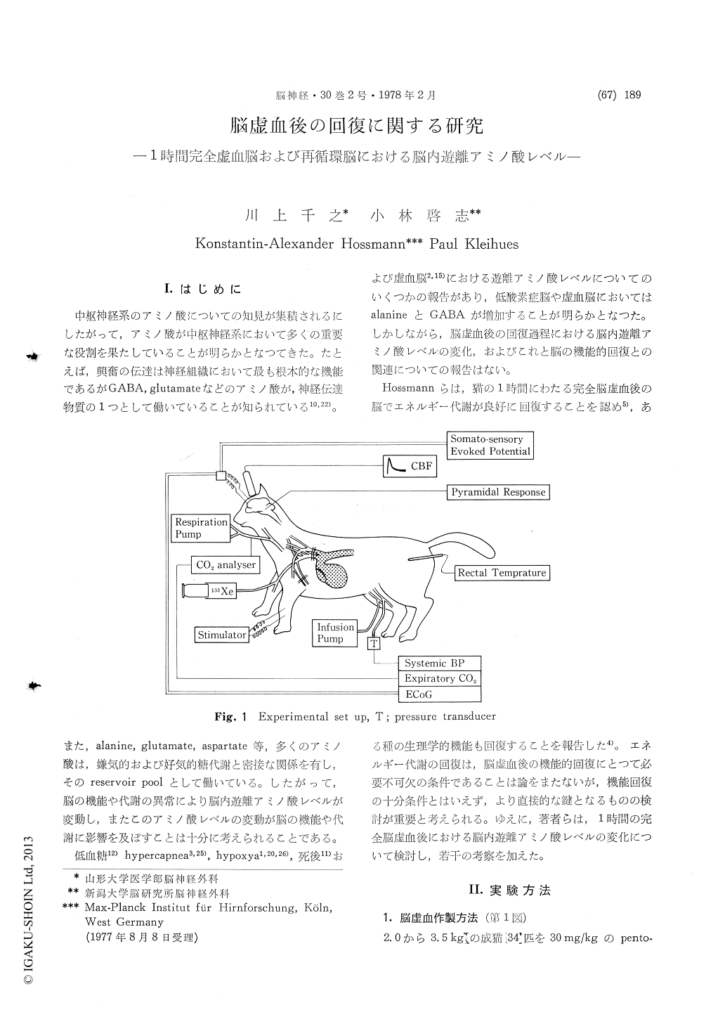
To clarify the mechanism of recovery after cere-bral ischemia, authors observed changes in free amino acid levels in the brain tissue during and following one hour complete cerebral ischemia in the cat. Adult cats were anesthetized with pentobarbital and immobilized with gallamine triethiodide, and artificially respirated. Left thoracotomy was done and the internal mammary arteries were ligated. The innominate and the left subclavian arteries were clamped immediatly after the blood pressure was reduced to 80 mmHg. Soon after the arterial clamping, EEG became flat and wash-out curve of 133Xe revieled completeness of the cerebral ischemia. Following one hour ischemia, blood pressure was reised and the arterial clamps were removed to initiate the recirculation. About three hours after the initiation of recirculation, the EEG reappeared.
Aspartate, asparagine, glutamate, glutamine, alanine and GABA levels in the brain tissue were measured. The brain tissue was taken and freeze-clamped in the liquid nitrogen as quickly as possible (within 2 or 3 seconds). The freeze-clamped material was homogenized, deproteinized and amino acid content was measured with liquid colomn chromatography.
The most pronounced change which developed during one hour ischemia was an increase in alanine followed by GABA, and glutamine. Asparagine and glutamate did not change significantly. Aspartate was decreased. During the early phase of recirculation (within 3 hours recirculation), alanine continued to increase tremendously, whereas glutamate and aspartate decreased. At longer re-circulation times, abnormal levels in alanine and glutamate tended to normalize. The noticeableevidence of them is that change in the free amino acid levels was most remarkable during the early phase of recirculation, not during the ischemia and it was normalized at longer times of recirculation. The changes during the ischemia was assumed to be due to arrest of the oxydative metabolism and that during the early recirculation phase due to compensate block in the pathway from the anearobic glycolysis to the TCA-cycle which started to function. In the late recirculation phase, as thecarbohydrate metabolism tended to return to normal so the amino acid levels tended to return to nomal.
It was shown that when the glutamate level and the GABA/glutamate ratio were abnormal, the EEG did not return, whereas they returned to normal the EEG began to reappear. This evidence indicates some relationship between these amino acid levels and recovery of brain function after ischemia.
Copyright © 1978, Igaku-Shoin Ltd. All rights reserved.