Japanese
English
- 有料閲覧
- Abstract 文献概要
- 1ページ目 Look Inside
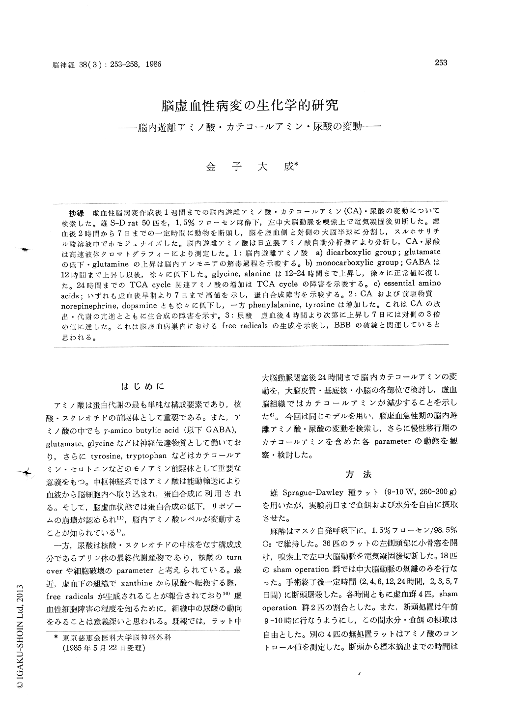
抄録 虚血性脳病変作成後1週間までの脳内遊離アミノ酸・カテコールアミン(CA)・尿酸の変動について検索した。雄S-D rat 50匹を,1.5%フローセン麻酔下,左中大脳動脈を嗅索上で電気凝固後切断した。虚血後2時間から7日までの一定時間に動物を断頭し,脳を虚血側と対側の大脳半球に分割し,スルホサリチル酸溶液中でホモジェナイズした。脳内遊離アミノ酸は日立製アミノ酸自動分析機により分析し,CA・尿酸は高速液体クロマトグラフィーにより測定した。1:脳内遊離アミノ酸 a) dicarboxylic group;glutamateの低下・glutamineの上昇は脳内アンモニアの解毒過程を示唆する。 b) monocarboxylic group;GABAは12時間まで上昇し以後,徐々に低下した。glycine, alanineは12-24時間まで上昇し,徐々に正常値に復した。24時間までのTCA cycle関連アミノ酸の増加はTCA cycleの障害を示唆する。 c) essential aminoacids;いずれも虚血後早期より7日まで高値を示し,蛋白合成障害を示唆する。2:CAおよび前駆物質norepinephrine, dopamineとも徐々に低下し,一方phenylalanine, tyrosineは増加した。これはCAの放出・代謝の亢進とともに生合成の障害を示す。3:尿酸 虚血後4時間より次第に上昇し7日には対側の3倍の値に達した。これは脳虚血病巣内におけるfree radicalsの生成を示唆し, BBBの破綻と関連していると思われる。
Changes in cerebral free amino acids, cate-cholamines and uric acid levels were explored for up to 7 days after cerebral ischemia in the rat. Fifty male Sprague-Dawley rats were subjected to occlusion of the middle cerebral artery on the olfactory tract, under halothane anesthesia.
The animals were decapitated at 2, 4, 6, 12, 24 hours and 2, 3, 5, 7 days after the surgery, respec-tively. The brains were rapidly removed. The cerebral hemispheres were divided into right and left halves, and homogenized in sulfosalicylic acid solution. Free amino acids were analyzed by color-metric method. Cathecholamines and uric acid were analyzed by high-performance liquid chro-matography. Each parameters were measured both on the ischemic and contralateral hemi-spheres. The time course of changes in each para-meters were observed by means of the ratio, which is the value of ischemic side divided by that of contralateral side.
1. Free amino acids a) Dicarboxylic group ; Decreases in glutamate and increases in glutamine suggest one aspect of detoxication of ammonia within the ischemia tissue. b) Monocarboxylic group ; GABA, glycine, alanine were increased in early ischemic state, and gradually lowered to the normal values. These suggest the impair-ment of tricarboxylic acid (TCA) cycle in the ischemic tissus, since these anino acids are closely related to TCA cycle. c ) Essential amino acids, except for tryptophan, were increased until the end of study. These increases suggest the utiliza-tion of essential amino acids for protein synthesis might be disturbed in the ischemic tissues. 2. Catecholamines and precursors ; Norepinephrine and dopamine were lowered gradually. On the other hand, phenylalanine and tyrosine were in-creased during ischemia. This result suggests the impaired biosynthesis of catecholamines followed by disutilization of phenylalanine and tyrosine in the ischemic tissue. 3. Uric acid increased gra-dually, reached to three-hold of control value at the end of study. It is known that free radicals are produced in the circuitry of uric acid syn-thesis. Then, this result suggests some degree of derangement in Blood-Brain-Barrier following the induction of free radical reaction in the infarcted lesion.

Copyright © 1986, Igaku-Shoin Ltd. All rights reserved.


