Japanese
English
- 有料閲覧
- Abstract 文献概要
- 1ページ目 Look Inside
I.はじめに
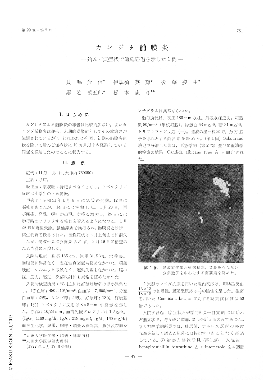
カンジダによる髄膜炎の報告は比較的少ない。またカンジダ髄膜炎は従来,末期的感染症としてその重篤さが強調されているが3),われわれは今回,初期の髄膜炎症状を除いて殆んど無症状に10カ月以上も経過している同症を経験したのでここに報告する。
An 11-year-old boy with Candida meningitis was reported. He was in excellent health until January 6, 1976, when he began to have high fever, headache and vomiting. A diagnosis of meningitis was made by a family physician who performed a spinal tap. Many kinds of antibiotics were administrated. About a month later, symptoms were subsided, but cerebrospinal fluid (CSF) still showed pleocytosis, elevation of protein and decreased sugar content.
He was admitted to the hospital on March 19, 1976. The physical examination on admission revealed no abnormalities. The CSF was under normal pressure, but pleocytosis (80/mm3, mononuclear cells), high protein (53 mg/dl) and lowered sugar content (31 mg/dl) were noted. Microscopic examination of CSF sediment with india ink revealed the presence of fungus elements. It was identified as Candida albicans type A by culture of CSF. Benzylpenicillin benzathine and clotrimazole were not effective. Two weeks after administration of 5-fluorocytosine, Candida was not found in the culture of CSF, but pleocytosis, elevation of protein and lowered sugar content persisted.
After the admission, deep reflexes became midly hyperactive bilaterally without pathological reflexes. Otherwise, no changes were seen physi-cally.
He had not taken antibiotics, ACTH or steroid hormone before the onset. He did not have under-lying disorders such as diabetes mellitus, leukemia or hypogammaglobulinemia. The route of invasion of fungus was unclear.
The present case showed that Candida meningitis may show a oligosymptomatic and prolonged course.

Copyright © 1977, Igaku-Shoin Ltd. All rights reserved.


