Japanese
English
- 有料閲覧
- Abstract 文献概要
- 1ページ目 Look Inside
I.はじめに
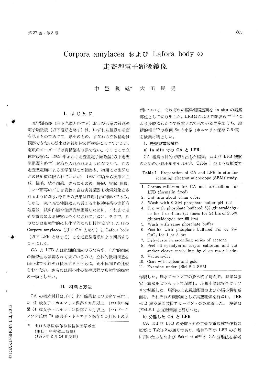
光学顕微鏡(以下光顕と略する)および通常の透過型電子顕微鏡(以下電顕と略す)は,いずれも組織の断面を見るものであつて,形そのもの,すなわち立体構造は観察できない。従来は連続切片の再構築によつていたが,電顕のオーダーでは再構築も容易でない。そこでこの立体的観察に,1962年頃から走査型電子顕微鏡(以下走査型電顕と略す)が取り入れられるようになつた2)。この走査型電顕による医学領域での観察も,初期には歯牙などの硬組維に限られていたが,1967年頃から次第に血球,繊毛,結合組織さらにその後,肝臓,腎臓,脾臓,リンパ節等のごとき管腔に富む実質臓器も検索対象とされるようになり,今日その成果は日進月歩の勢いである。しかし,完全充実性臓器とも言える中枢神経系の実質内観察は,試料作製や像解析が困難なために,これまで走査型電顕による観察は全くなされていない。そこで,このたびは形態学的にも化学的にも比較的安定した形のCorpora amylacea (以下CAと略す)とLafora body(以下LFBと略する)とを走査型電顕により観察することにした。
CAとLFBとは電顕的組成のみならず,化学的組成の類似性も強調されて来ているので,立体的微細構造を両小体でそれぞれ検索するとともに,両小体間での比較をおこない,さらには両小体の発生過程の形態学的探索の一助としたい。
The stereostructures of corpora amylacea (CA) in the corpus callosum and the Lafora body (LFB) in the cerebellum have been investigated with a scanning electron microscope. The specimens of CA and LFB were isolated materials and paraffin sections from these tissues, and only the subepen-dymal surface for CA.
CA was found to be an entirely full, homogeneous and very dense sphere, and the external surface constituted granular undulation of 35-130 mμ, in various widths. The external surface was partly combined closely with bundles of the glial fibrils.
LFB consisted of double structures which were internal and external layers. The internal layer was a full, homogeneous and very dense sphere, and was similar to CA. This layer shifted conti-nuously to the external layer which was composed of cables of 350 mμ-2μ, in various widths. The cables congregated rosarily in numerous spherules of 150-450 mμ in diameter and/or some granules of 35-70 mp in diameter.
Because of the stereostructural resemblances bet-ween CA and the internal layer of LFB and the similarities of each ultrastructural unit and chemical composition, it was suggested that these bodies were formed by similar mechanisms and that some of structural and chemical differences were due to the peculiarities of the cells and tissues in which these bodies were produced.

Copyright © 1975, Igaku-Shoin Ltd. All rights reserved.


