Japanese
English
- 有料閲覧
- Abstract 文献概要
- 1ページ目 Look Inside
I.はじめに
近年,不随意運動症と生体アミンの変動,ことにpa—rkinsonismにおけるdehydroxyphenyl-alamine, dopa—mine系の代謝障害が注目され,またdopamineは尾状核に特に多いことが明らかにされているが,われわれは今回,定位脳手術を行なつたparkinsonism及び痙性斜頸や舞踏病など(hyperkinetic group)の症例において手術前の気脳撮影前後像上より想定される尾状核頭部の大きさの変化と臨床症状とを対比し,検討を試みた。
Dividing the patients with involuntary move-ments into parkinsonism group and hyperkinetic group, we observed the size of the head of caudate nucleus on A-P projection in PEG, in relation to clinical symptoms.
We measured the followings ;
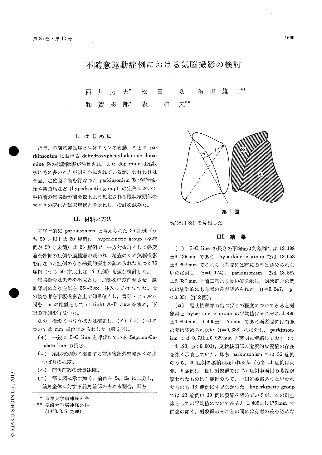
(イ) The length of Septum-Caudate line.
(ロ) The convex part of the head of the cau- date nucleus.
(ハ) The longest distance of frontal horn.
(ニ) S1/S1+S2
(S1; posterior part of frontal horn.)
(S2; anterior part of frontal horn.)
In parkinsonism there was a selective dilatation of the posterior part of the frontal horn, which means an atrophy of the head of the caudate nucleus. This change was particularly marked in the patients of the age over 50.
Patients with parkinsonism were further divided into rigidity group and tremor group. The degree of atrophy of the head of the caudate nucleus was more marked in rigidity group.
On the other hand, the ventricular system was generally dilated in tremor group. No marked difference was noted between the affected and the normal sides.
In hyperkinetic group, comparing the control group, marked differences were recognized in the length of Septum-Caudate line and in the convex part of the head of the caudate nucleus.
However, the hyperkinetic group is longer in the longest distance of the frontal horn and bigger inS1/S1+S2 In this group, selective dilatation in anterior part of frontal horn was noted.

Copyright © 1973, Igaku-Shoin Ltd. All rights reserved.


