Japanese
English
- 有料閲覧
- Abstract 文献概要
- 1ページ目 Look Inside
緒言
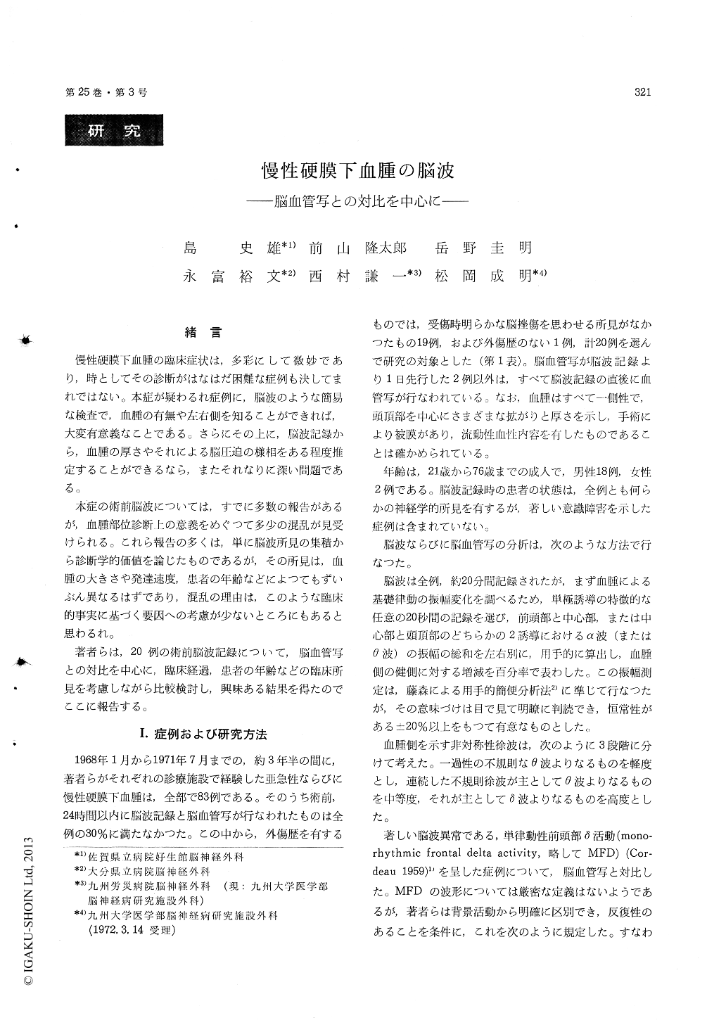
慢性硬膜下血腫の臨床症状は,多彩にして微妙であり,時としてその診断がはなはだ困難な症例も決してまれではない。本症が疑わるれ症例に,脳波のような簡易な検査で,血腫の有無や左右側を知ることができれば,大変有意義なことである。さらにその上に,脳波記録から,血腫の厚さやそれによる脳圧迫の様相をある程度推定することができるなら,またそれなりに深い問題である。
本症の術前脳波については,すでに多数の報告があるが,血腫部位診断上の意義をめぐつて多少の混乱が見受けられる。これら報告の多くは,単に脳波所見の集積から診断学的価値を論じたものであるが,その所見は,血腫の大きさや発達速度,患者の年齢などによつてもずいぶん異なるはずであり,混乱の理由は,このような臨床的事実に基づく要因への考慮が少ないところにもあると思わるれ。
In 20 cases of unilateral chronic subdural he-matoma the EEG findings were reviewed in relation to the cerebral angiography and some other clinical findings. The results were as follows.
1) Decreased amplitude of alpha activity was found in cases where the hematoma developed rapidly after the neurological symptoms had appeared. But there was no relation found be-tween the thickness of the hematoma and the de-crease of the amplitude.
2) Increased amplitude of the alpha activity wasalways associated with a thick hematoma, which had developed slowly during a long clinical term.
3) Marked asymmetric slow waves on the side of the hematoma were seen in cases with a thick hematoma, which had developed rapidly after the manifestation of neurological symptoms.
4) Monorhythmic frontal delta activity was found in cases which in the angiogram showed a remark-able shift of the anterior cerebral artery.
5) Slow alpha activity due to the existence of a hematoma was found only in elderly patients.
As seen above, in cases of subdural hematoma, the EEG findings had some relation to the thick-ness of the hematoma and the shift of the anterior cerebral artery and also were depending consider-ablly on the development of the clinical course of the hematoma.
In two cases the EEG recordings showed no ab-normal findings, in spite of the fact that hemato-mas actually existed. It should be noted that these hematomas were not smaller in size than those of the other cases. This again indicates that in cases of subdural hematoma, the EEG recordings are not always reliable.

Copyright © 1973, Igaku-Shoin Ltd. All rights reserved.


