Japanese
English
- 有料閲覧
- Abstract 文献概要
- 1ページ目 Look Inside
硬膜下血腫の脳波に関しては,Jasper1)以来,Williams2),桂3),本川4),Cohn5),Walter6),Schwab7),等により,多くの詳細なる研究がなされて来て居る。然し,一定の見解はなく,血腫領野に局在した徐波,低電位,又血腫の部位,大きさ等の相違により,それが皮質に与える影響の如何によつて,脳波の変化は多様であり,Walter等は正常脳波を示すものもあると報告している。
桂外科教室に於ては,亜急性,或は慢性硬膜下血腫,31例を経験して居るが,其の中,16例について脳波による検索を行つて居り,其の総合的観察の結果,2〜3の興味ある所見を得たので報告する。
Of the 31 cases with subdural hematoma treated at the Surgical Clinic of Prof. S. Ka-tsura, Tohoku University School of Medicine, 16 cases were subjected to EEG examination, for studies from various angles.
Generally speaking, the EEG of such sub-dural hematoma shows so little anomaly that the changes are easily overlooked, but all the 16 cases examined in this study showed perceptible changes in some way.
The EEG findings most serviceable in in-dicating the site of the tumor consisted in local foci of inhibited or slackened brain waves, which could he detected homolateral-ly in most cases. It is to be noted, however, that in some cases such foci were found contralaterally, so that the localized inhibi-tion and retardation foci may be very ser-viceable findings offering basis for diagnos-ing the location of the hematoma, but cannot be called 100% precise indicators.
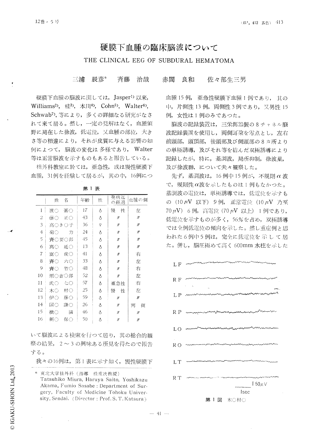
The fundamental EEG showed in all cases irregular a-waves as the dominant waves and the potential tended to be low, especially among the cases with advanced tumors. In only one case with high intercranial pressure, however, the potential was found supernor-mal.

Copyright © 1960, Igaku-Shoin Ltd. All rights reserved.


