Japanese
English
- 有料閲覧
- Abstract 文献概要
- 1ページ目 Look Inside
I.序論
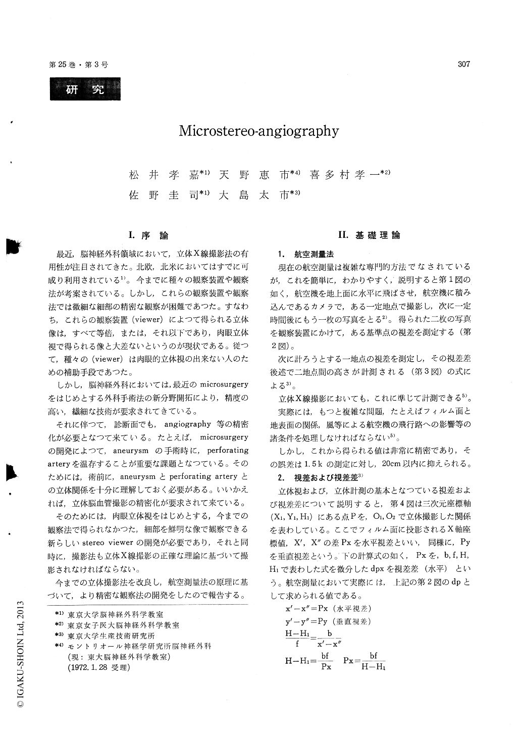
最近,脳神経外科領域において,立体X線撮影法の有用性が注目されてきた。北欧,北米においてはすでに可成り利用されている1)。今までに種々の観察装置や観察法が考案されている。しかし,これらの観察装置や観察法では微細な細部の精密な観察が困難であつた。すなわち,これらの観察装置(viewer)によって得られる立体像は,すべて等倍,または,それ以下であり,肉眼立体視で得られる像と大差ないというのが現状である。従って,種々の(viewer)は肉眼的立体視の出来ない人のための補助手段であつた。
しかし,脳神経外科においては,最近のmicrosurgeryをはじめとする外科手術法の新分野開拓により,精度の高い,繊細な技術が要求されてきている。
Recent advance in roentgen-stereography has clarified definite importance of precise preoperative assessment of stereoscopic X ray films toward diagnosis and treatment of intracranial disorders. This report is concerned with a new development in stereoradiology using specifically designed stereo-viewer for precise evaluation of cerebral angio-graphy. Microscopic dissection of intracranial arte-riovenous malformations and aneurysms requires special operative care not to jeopardize small per-forating vessels around the aneurysms, which neces-sitates well-organized radiological system to establish three-dimensional approach to the above-mentionedmorbid anatomy. Previous techniques on stereo-scopic radiology are extensively reviewed and criti-cized to improve the quality of stereoscopic films. Conventional stereoscopic films have been unable to give us precise three-dimensional arrangement of small intracranial vessels in close geographical approximation. Based on the knowledge and ex-perience of aerial photography, the most appropriate conditions to take stereoscopic X ray films are precisely calculated. Distance of the parallel move-ment of the X ray tube is determined for micro-surgical dissection of the intracranial aneurysms and nearby vascular structures as well as for routine stereoscopic X ray films. A new stereoviewer is designed in cooperation with Nikon Co. Ltd. This viewer is used with adequate magnification in the office and valuable in the operating room together with stereoscopic films taken in the direction of any desired craniotomy to give us three-dimensional arrangement of intracranial vascular structures at the time of actual microsurgical dissection. By superimposing several different stereoscopic films, it is possible to obtain both arterial and venous phases in one stereoscopic vision. A new Lysholm Blende is also introduced with special grid ratio for stereoscopic films to obtain better contrast of the films. Together with our new techniques on measuring various geographical distances among intracranial vasculature, particularly small perforat-ing vessels and arterial feeders of arteriovenous malformations, the possibility of computer analysis on cerebral angiography is discussed.

Copyright © 1973, Igaku-Shoin Ltd. All rights reserved.


