Japanese
English
- 有料閲覧
- Abstract 文献概要
- 1ページ目 Look Inside
I.はじめに
Sturge-Weber病はその臨床的特徴から主として皮膚科,眼科,精神科領域で取り扱われてきたが,すでに1879年Sturge1)が脳病変の存在を予言し,1935年Moniz8)以来脳内血管の造影により本疾患の脳内変化に対する探索14)16)がなされてきた。しかし広汎な脳血管腫様変化のために積極的な外科的治療は行なわれにくく,したがつて手術経験例16)28)29)30)も文献上少ないように思われる。一方頭蓋内に発生するPlexuspapillomaは統計6)17)25)上かなりまれな脳腫瘍であり,本邦報告21)26)31)例も散見されるにすぎない。
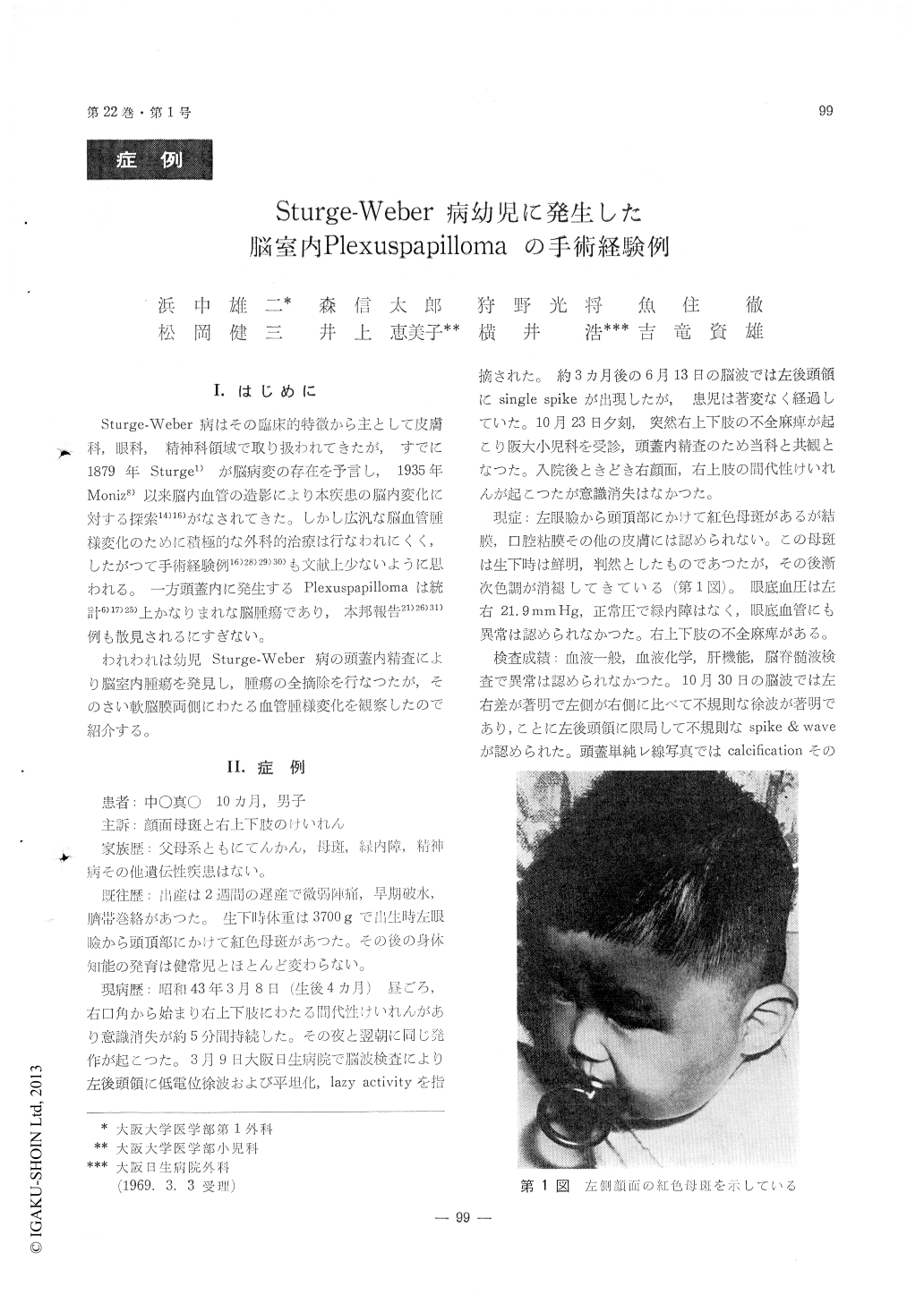
われわれは幼児Sturge-Weber病の頭蓋内精査により脳室内腫瘍を発見し,腫瘍の全摘除を行なつたが,そのさい軟脳膜両側にわたる血管腫様変化を観察したので紹介する。
A ten-month-old boy was reported who was noted a port-wine nevi on the left side of the forehead at birth. At four months of age, he had convulsions of the right arm and leg, and admitted to our hospital at ten months.
On examination, hemiparesis of the right arm and leg were recongnized. An electroencephalogram showed spike and wave-complex on the left occipital region. Intracranial calcification was not visible on x-ray film, but cortical atrophy was suggested by cerebral angiography and a round tumor shadow at the trigone of the left lateral ventricle by pneu-moencephalography.
When the right ventricular drainage and the left temporo-parieto-occipital craniotomy were done, venous angioma of the leptomeninges were revealed. After the opening of the left ventricle, a round red-dish tumor was found and totally extirpated.
Histologically the tumor was a typical papilloma of the choroid plexus. No malignant cells were found.
The patient was discharged without convulsion.

Copyright © 1970, Igaku-Shoin Ltd. All rights reserved.


