Japanese
English
- 有料閲覧
- Abstract 文献概要
- 1ページ目 Look Inside
I.緒言
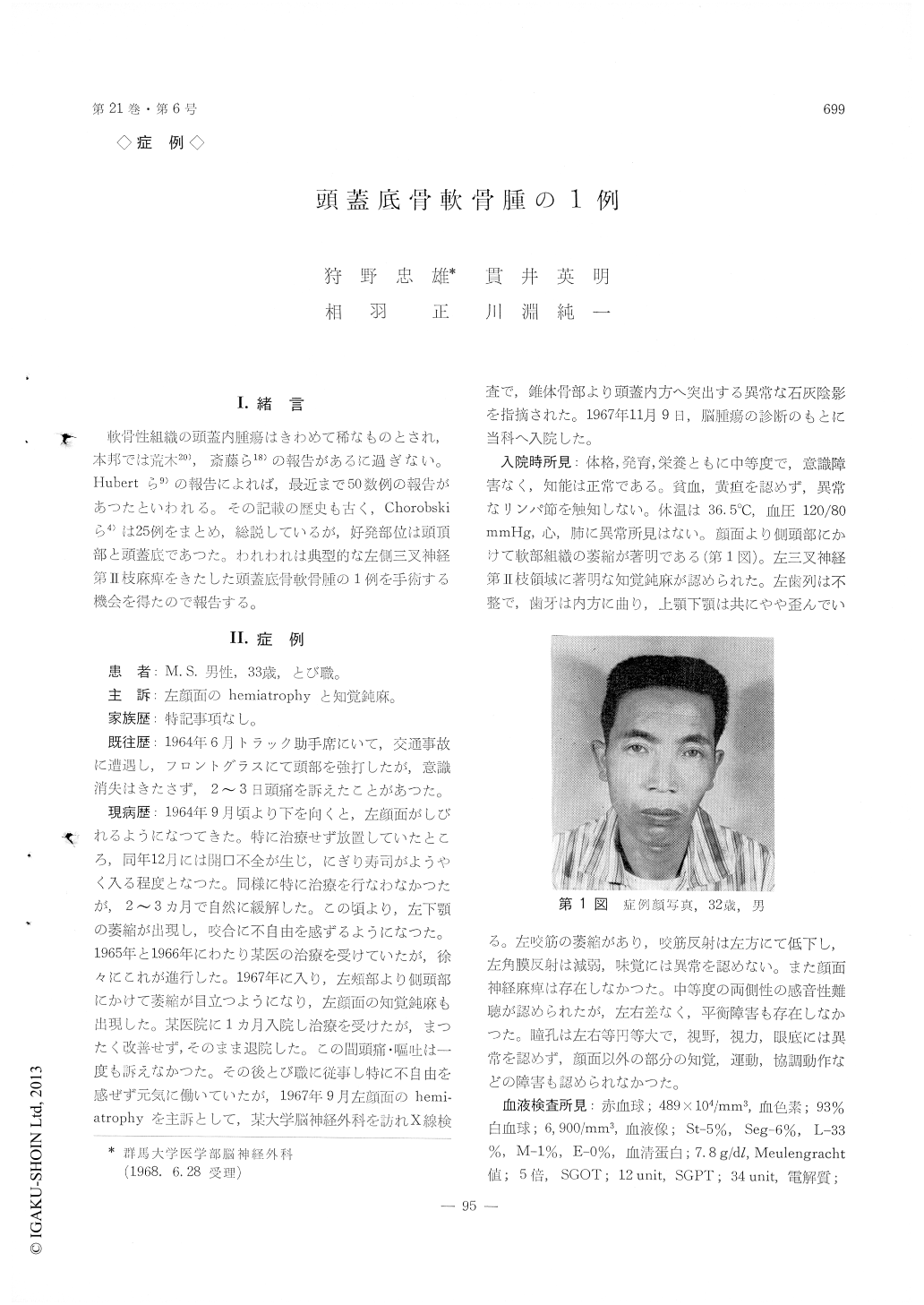
軟骨性組織の頭蓋内腫瘍はきわめて稀なものとされ,本邦では荒木20),斎藤ら18)の報告があるに過ぎない。Hubertら9)の報告によれば,最近まで50数例の報告があつたといわれる,その記載の歴史も古く,Chorobskiら4)は25例をまとめ,総説しているが,好発部位は頭頂部と頭蓋底であつた。われわれは典型的な左側三叉神経第II枝麻痺をきたした頭蓋底骨軟骨腫の1例を手術する機会を得たので報告する。
The rarity of intracranial chondroma is demons-trated by the fact that Cushing found only three chondromas among 2023 brain tumors and Tonnis classified nine chondromatous tumors in his series of 4135 brain tumors. Since Hirschfeld had reported the first case, over fifty cases have been hitherto pulished. Such lesion may arise from the base of the skull or parietal region.
A case of primary osteochondroma in the left middle cranial fossa is reported. On Nov. 9, 1967, a 32-year-old male patient was admitted to the Gunma University Hospital with a three year history of left hemiatrophy of the face, characterized by the paresis of trigeminal nerve. The Roentgen studies revealed that the small hen egg-sized and calcified tumor occupied the middle cranial fossa and destructed the tip of the pyramidal bone. The sub-total surgical excision was carried out with some difficulties on Nov.30, 1967. The tumor was located in the extradural space of the middle cranial fossa and extended from the parasellar region to the py-ramidal hone destructing the bony structures in this region.
The pathological examinations disclosed that chon-dromatous cells were scattered in the cartilage ma-trix and trabeculae of hone were found in some places of the tumor tissue. The diagnosis of os-teochondroma was made to this tissue.
On Dec. 19, 1967, the patient was discharged with paresis of the left trigeminal nerve. He is now working daily in good health.

Copyright © 1969, Igaku-Shoin Ltd. All rights reserved.


